. 2007 Mar;13(3):396-403.
doi: 10.3201/eid1303.060549.

In vitro cell culture infectivity assay for human noroviruses

Affiliations

In vitro cell culture infectivity assay for human noroviruses

Timothy M Straub et al. Emerg Infect Dis. 2007 Mar.

Abstract

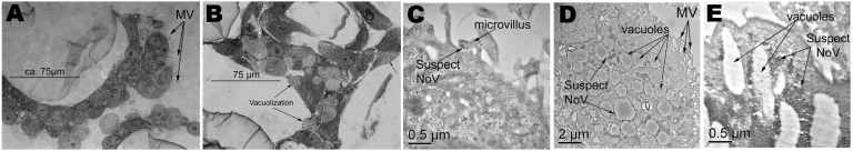
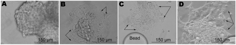
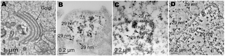
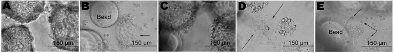
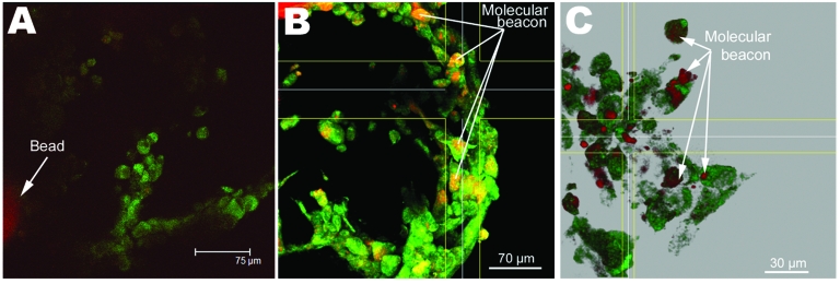
Human noroviruses cause severe, self-limiting gastroenteritis that typically lasts 24-48 hours. Because of the lack of suitable tissue culture or animal models, the true nature of norovirus pathogenesis remains unknown. We show, for the first time, that noroviruses can infect and replicate in a physiologically relevant 3-dimensional (3-D), organoid model of human small intestinal epithelium. This level of cellular differentiation was achieved by growing the cells on porous collagen-I coated microcarrier beads under conditions of physiological fluid shear in rotating wall vessel bioreactors. Microscopy, PCR, and fluorescent in situ hybridization provided evidence of norovirus infection. Cytopathic effect and norovirus RNA were detected at each of the 5 cell passages for genogroup I and II viruses. Our results demonstrate that the highly differentiated 3-D cell culture model can support the natural growth of human noroviruses, whereas previous attempts that used differentiated monolayer cultures failed.


Figures

Figure 1
Figure 1
Light and transmission electron micrographs of uninfected and infected tissue aggregates with a combined stock of noroviruses representing 3 strains (Passage 0 [P0]). A) Uninfected tissue aggregates displaying well-formed microvilli. B) Infected tissue aggregates exhibiting vacuolization and shortening of the microvilli. C) Transmission electron microscopy (TEM) at 1 h postinfection showing possible norovirus in a microvillus. D) TEM at 24 h postinfection showing significant vacuolization, and internal membrane rearrangement. E) TEM at 66 h postinfection showing accumulation of suspect norovirus particles.
Figure 2
Figure 2
Demonstration of cytopathic effect in infected tissue aggregates during the second infection trial. A) Uninfected aggregate, 24 h into the experiment. B) Cells infected with lysate from the first infection trial (P1) at 24 h postinfection. C) Stool sample flag2 at 24 s postinfection (P0). D) Stool sample 149 at 48 h postinfection (P0). Arrows indicate cells exhibiting unusual pathology.
Figure 3
Figure 3
Transmission electron microscopy of uninfected and infected cell cultures from the second infection trial. A) Uninfected cells showing normal internal membrane organelles. B) Suspect 29-nm particles in cells, viruses from cell culture lysate from the first infection trial (P1). C) Stool sample flag2 (P0) and D) stool sample 149 (P0) showing numerous 29-nm particles and internal rearrangement of membrane-bound organelles.
Figure 4
Figure 4
Cytopathic effect results from the third infectivity trial. A) Virus-free control of B) combined viral stock lysate from second passage experiment (second infectivity trial, P1), which was used to infect naive cells (P2). C) Virus-free control of the flag2 stool sample. D) Corresponding infection with the flag2 stool sample (P0). E) Flag2 in cell culture (P1). Cells in Panels B, D, and E were confirmed as positive for norovirus by reverse transcription–PCR (RT-PCR) and seminested PCR. Cells in uninfected controls were negative for norovirus by both RT-PCR and nested PCR. Arrows indicate cells exhibiting unusual pathology.
Figure 5
Figure 5
Deconvolved confocal laser scanning micrographs of the molecular beacon fluorescence in situ hybridization assay, demonstrating viral infectivity of a genogroup I virus (Sample 155) and genogroup II virus (flag2). A) Typical response for uninfected cells, no molecular beacon observed. B) Sample 155, P5 in cell culture. C) Sample flag2, P5 in cell culture.

Comment in

  • Cell culture assay for human noroviruses.
    Chan MC, Wong YP, Leung WK. Chan MC, et al. Emerg Infect Dis. 2007 Jul;13(7):1117; author reply 1117-8. doi: 10.3201/eid1307.070131. Emerg Infect Dis. 2007. PMID: 18214197 Free PMC article. No abstract available.

Similar articles

Cited by

References

    1. Atmar RL, Estes MK. Diagnosis of noncultivatable gastroenteritis viruses, the human caliciviruses. Clin Microbiol Rev. 2001;14:15–37. 10.1128/CMR.14.1.15-37.2001 - DOI - PMC - PubMed
    1. de Wit MA, Koopmans MP, Kortbeek LM, Wannet WJ, Vinje J, van Leusden F, et al. Sensor, a population-based cohort study on gastroenteritis in the Netherlands: incidence and etiology. Am J Epidemiol. 2001;154:666–74. 10.1093/aje/154.7.666 - DOI - PubMed
    1. Hale A, Mattick K, Lewis D, Estes M, Jiang X, Green J, et al. Distinct epidemiological patterns of Norwalk-like virus infection. J Med Virol. 2000;62:99–103. 10.1002/1096-9071(200009)62:1<99::AID-JMV15>3.0.CO;2-0 - DOI - PubMed
    1. Lindesmith L, Moe C, Marionneau S, Ruvoen N, Jiang X, Lindblad L, et al. Human susceptibility and resistance to Norwalk virus infection. Nat Med. 2003;9:548–53. 10.1038/nm860 - DOI - PubMed
    1. Parashar U, Quiroz ES, Mounts AW, Monroe SS, Fankhauser RL, Ando T, et al. “Norwalk-like viruses.” Public health consequences and outbreak management. MMWR Recomm Rep. 2001;50(RR-9):1–17. - PubMed

Publication types

LinkOut - more resources