Review
. 2007 Aug;211(2):188-98.
doi: 10.1111/j.1469-7580.2007.00750.x. Epub 2007 Jun 6.

Slit-Robo interactions during cortical development

Affiliations
Review

Slit-Robo interactions during cortical development

William D Andrews et al. J Anat. 2007 Aug.

Abstract

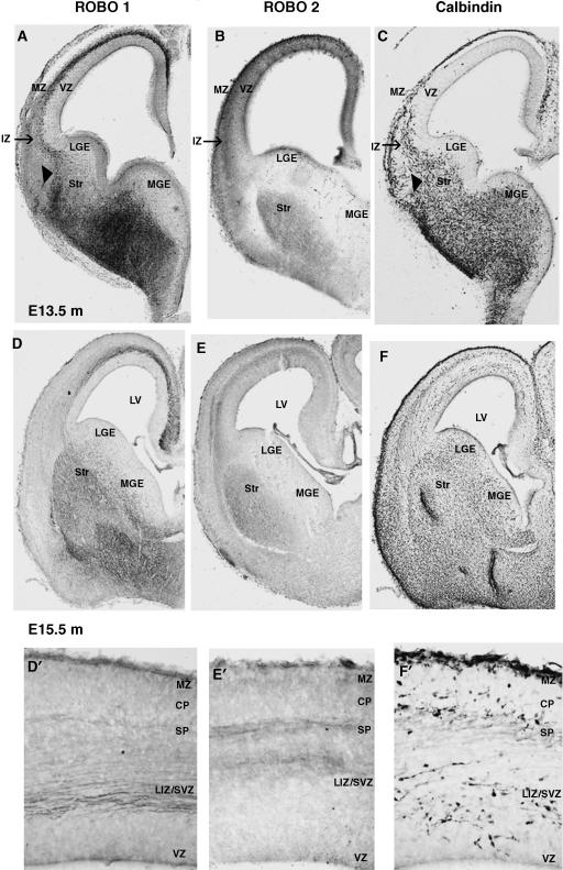
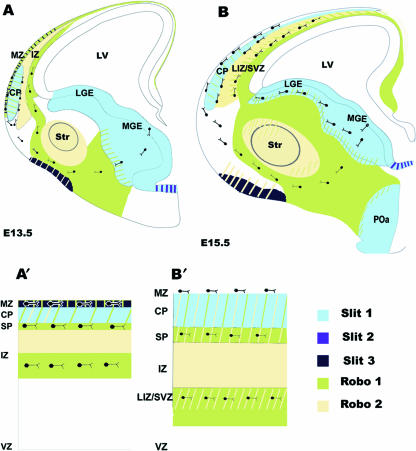
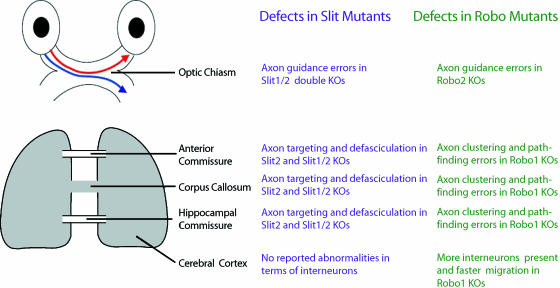
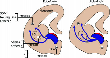
Interneurons are an integral part of cortical neuronal circuits. During the past decade, numerous studies have shown that these cells, unlike their pyramidal counterparts that are derived from the neuroepithelium along the lumen of the lateral ventricles, are generated in the ganglionic eminences in the subpallium. They use tangential migratory paths to reach the cortex, guided by intrinsic and extrinsic cues. Evidence is now emerging which suggests that the family of Slit proteins, acting through Robo receptors, play a role not only in axon guidance in the developing forebrain, but also as guiding signals in the migration of cortical interneurons. Here we describe the patterns of expression of Slit and Robo at different stages of forebrain development and review the evidence in support of their role in cortical interneuron migration. Slit-Robo signal transduction mechanisms are also important during normal development in a number of systems in the body and in disease states, making them potential therapeutic targets for the treatment of neurological disorders and certain types of cancer.


Figures

Fig. 1
Fig. 1
Expression patterns of Robo proteins during cortical development. Images illustrate the immunohistochemical localization of Robo1, Robo2 and Calbindin proteins in coronal sections through the developing mouse forebrain at E13.5 (A–C) and E15.5 (D–F). Enlarged portions of the dorsal cortex at E15.5 are shown for Robo1 (D′), Robo 2 (E′) and Calbindin (F′) localization. MZ, marginal zone; IZ, intermediate zone; VZ, ventricular zone; LGE, lateral ganglionic eminence; MGE, medial ganglionic eminence; Str, striatum; LV, lateral ventricle; CP, cortical plate; SP, subplate; LIZ/SVZ, lower intermediate zone/subventricular zone.
Fig. 2
Fig. 2
Schematic representation of Robo and Slit localization in the developing forebrain. Schematic drawings, based on in situ and immunohistochemical studies, illustrate robo and slit expression patterns during early (E13.5) (A) and mid (E15.5) (B) phases of cortical interneuron migration. Hatched patterns indicate regions of overlap in expression. Enlarged portions of the dorsal cortex at E13.5 (A′) and E15.5 (B′) show the localization of robo and slit and the regions of overlapping expression in greater detail. MZ, marginal zone; IZ, intermediate zone; VZ, ventricular zone; LGE, lateral ganglionic eminence; MGE, medial ganglionic eminence; Str, striatum; LV, lateral ventricle; CP, cortical plate; SP, subplate; LIZ/SVZ, lower intermediate zone/subventricular zone; POa, preoptic area.
Fig. 3
Fig. 3
Different developmental abnormalities observed in Slit and Robo null mice. Different phenotypical manifestations have been reported between Slit and Robo1 null mice in several systems. In both types of mutants an ectopic chiasm is observed at the optic chiasm, but only in slit mutants do we observe a dorsally projecting axon into the controlateral optic tract (Plump et al. 2002). In cortical commissures and at the corpus callosum Slit mutants demonstrate defects in axon targeting and defasciculation (Bagri et al. 2002; Whitford et al. 2002; Shu et al. 2003), whereas Robo mutants show axon pathfinding errors and axon clustering (Andrews et al. 2006).
Fig. 4
Fig. 4
Mechanism regulating migration of interneurons from the subpalium to the cerebral cortex in Robo1 wild-type (+/+) and Robo1 null (–/–) mice. Schematic drawings of coronal sections through the mouse telencephalon illustrate how in Robo1 wild-type (+/+) mice cortical inteneurons, generated in the subventricular zone of the medial ganglionic eminence (shown in blue), are expelled from this region by the repulsive action of Slit (shown in red). Unidentified repulsive activity (minus sign) present in the preoptic area (POa) prevents the migration of these neurons ventrally. Expression of Semaphorins in the developing striatum (Str) prevents cortical interneurons that express neuropilin and Robo from entering this structure in mice. Attractive factors (plus sign) including SDF-1 and neuregulins guide interneurons towards the cortex. However, in Robo1–/– mice, interneurons invade the striatal region ignoring the repulsive effects of Semaphorins and other factors, and enter the cortex earlier and in greater numbers than in wild-type littermates.

References

    1. Anderson SA, Eisenstat DD, Shi L, Rubenstein JL. Interneuron migration from basal forebrain to neocortex: dependence on Dlx genes. Science. 1997;278:474–476. - PubMed
    1. Andrews W, Liapi A, Plachez A. Robo1 regulates the development of major axon tracts and interneuron migration in the forebrain. Development. 2006;133:2243–2252. - PubMed
    1. Bagri A, Marín O, Plump AS. Slit prevents midline crossing and determines the dorsoventral position of major axonal pathways in the mammalian forebrain. Neuron. 2002;33:233–248. - PubMed
    1. Bashaw GJ, Kidd T, Murray D, Pawson T, Goodman CS. Repulsion axon guidance: Abelson and Enabled play opposing roles downstream of the roundabout receptor. Cell. 2000;101:703–715. - PubMed
    1. Battye R, Stevens A, Jacobs JR. Axon repulsion from the midline of the Drosophila CNS requires slit function. Development. 1999;126:2475–2481. - PubMed

Publication types

LinkOut - more resources