. 2007 Jun 7;54(5):697-711.
doi: 10.1016/j.neuron.2007.05.016.

Human medial frontal cortex mediates unconscious inhibition of voluntary action

Affiliations

Human medial frontal cortex mediates unconscious inhibition of voluntary action

Petroc Sumner et al. Neuron. .

Abstract

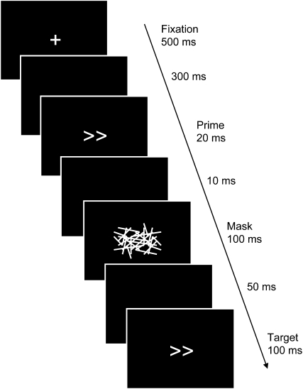
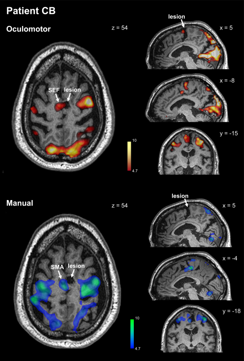
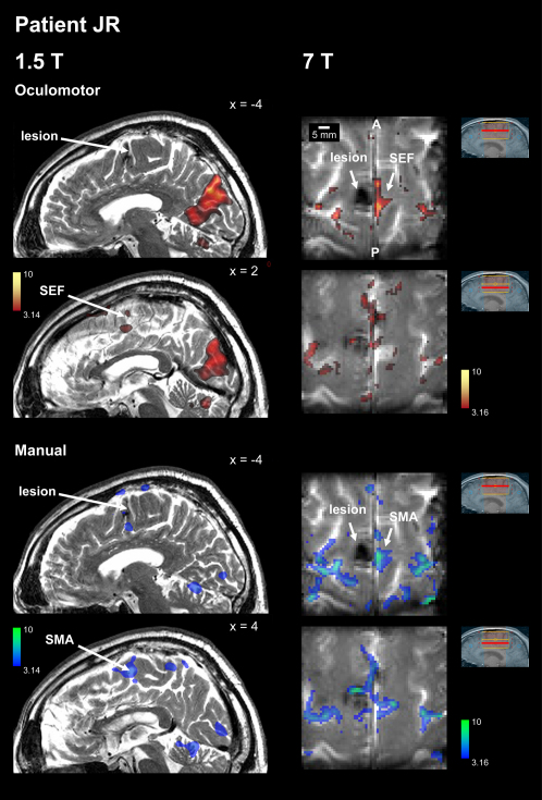
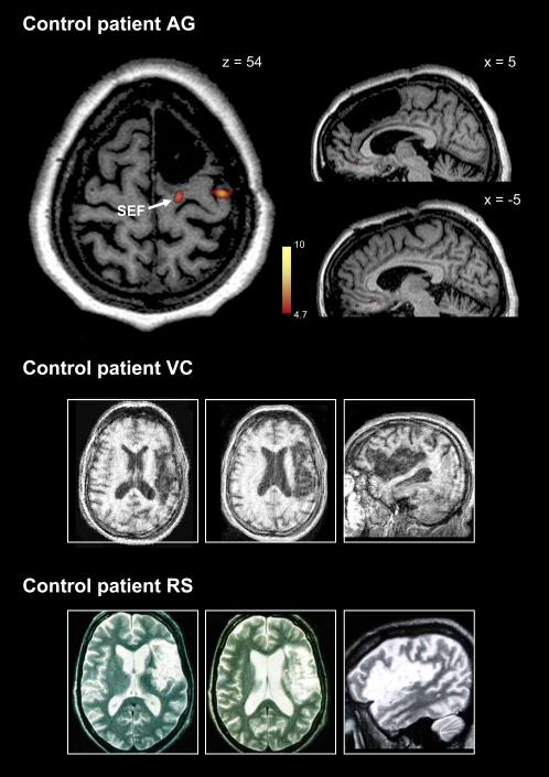
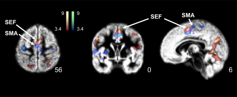
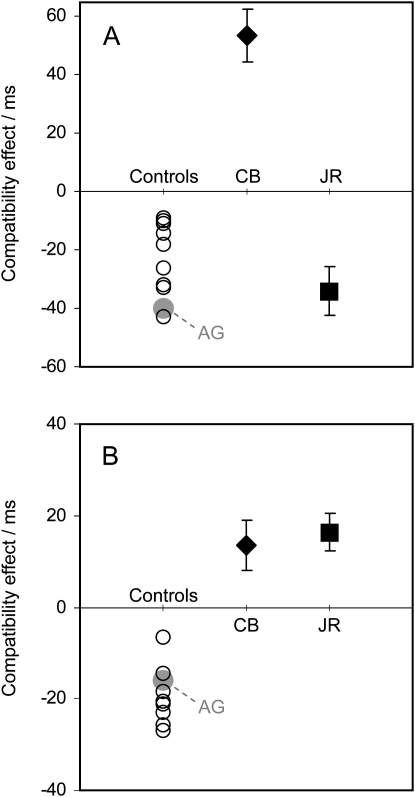
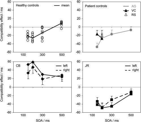
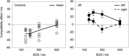
Within the medial frontal cortex, the supplementary eye field (SEF), supplementary motor area (SMA), and pre-SMA have been implicated in the control of voluntary action, especially during motor sequences or tasks involving rapid choices between competing response plans. However, the precise roles of these areas remain controversial. Here, we study two extremely rare patients with microlesions of the SEF and SMA to demonstrate that these areas are critically involved in unconscious and involuntary motor control. We employed masked-prime stimuli that evoked automatic inhibition in healthy people and control patients with lateral premotor or pre-SMA damage. In contrast, our SEF/SMA patients showed a complete reversal of the normal inhibitory effect--ocular or manual--corresponding to the functional subregion lesioned. These findings imply that the SEF and SMA mediate automatic effector-specific suppression of motor plans. This automatic mechanism may contribute to the participation of these areas in the voluntary control of action.


Figures

Figure 1
Figure 1
Illustration of the Stimulus Sequence in the Masked-Prime Task The participants responded to the target arrows, which could point left or right, by pressing left or right buttons. The prime, which could also point left or right, was rendered invisible by the mask, so that participants saw only the fixation cross, the mask, and the target. If the prime was the same as the target, as depicted, it is a “compatible” trial. If the prime points in the opposite direction, it is an “incompatible” trial. Note that following previous convention (e.g., Eimer and Schlaghecken [2003]), the trial's SOA is measured from the start of the mask until the start of the target: in this case 150 ms.
Figure 2
Figure 2
Functional Localization of CB's Lesion Statistical contrasts (thresholded at p < 0.001 uncorrected) for oculomotor activity (upper panels) or manual activity (lower panels) are superimposed on a T1-weighted anatomical scan. Oculomotor activity within the caudal SFG constitutes the functionally defined SEF and appears in the left hemisphere directly opposite the lesion in the right hemisphere. Likewise, manual activity within the caudal SFG constitutes the functionally defined SMA and appears opposite and caudal to the lesion. See text for more details. See Supplemental Data for clinical information and imaging methodology.
Figure 3
Figure 3
Functional Localization of JR's Lesion Functional images for oculomotor activity (upper panels) or manual activity (lower panels) were acquired at 1.5T (left panels) and at 7T (right panels). For 1.5T, statistical contrasts (thresholded at p < 0.001 uncorrected) are superimposed on a T2-weighted sagittal image acquired with resolution 1.6 × 0.575 × 0.575 mm. The high-resolution 7T functional maps (1 × 1 × 3 mm) are superimposed on the mean echoplanar image. The scale bars indicate the very small size of the lesion (note also that there is likely to have been signal dropout around the true lesion due to hemosiderin deposition). There is clear contralesional oculomotor activity precisely opposite the lesion, indicating that the ipsilesional SEF is damaged. The extent of damage to the SMA is less clear but is likely to be less than to the SEF, given the rostral position and small size of the lesion. See text for more details. See Supplemental Data for clinical information and imaging methodology.
Figure 4
Figure 4
Control Patients' Lesions Functional imaging for oculomotor activity in patient AG (left panel) demonstrates SEF activation in the lesioned hemisphere caudal to the lesion. Thus pre-SMA is damaged, but not SEF and SMA. Sagittal structural images to either side of midline (right panels) reveal the extent of surgical resection in AG. Patient VC and patient RS have extensive strokes involving lateral frontal cortex, including ventral and dorsal premotor regions, but no involvement of medial frontal cortex. See Supplemental Data for clinical information and imaging methodology.
Figure 5
Figure 5
Functional Localization of SEF and SMA in Healthy Participants Oculomotor activity (red-yellow) occurred reliably just rostral to manual activity (blue-green) in the SFG, and activity was confluent across the medial wall of the SFG. Group data (n = 10) are shown; see Supplemental Data for individual data and imaging methods.
Figure 6
Figure 6
Results for Manual and Saccadic Responses The compatibility effect is the difference between response time on incompatible and compatible trials (and error bars are the standard error of this difference). For all control subjects, there was a negative compatibility effect—i.e., responses were longer on compatible trials—for both manual (A) and saccadic (B) responses, indexing inhibition (open circles for neurologically healthy controls; gray circle for AG, the lesion control patient). CB, however, showed a strong positive compatibility effect, indicating that the inhibitory process is disrupted for both manual and saccade responses. JR, whose smaller lesion appears to largely spare SMA, showed a normal negative effect for manual responses (A) but a positive effect for saccadic responses (B). See text for more details.
Figure 7
Figure 7
Results for Manual Responses with Varying Prime-Target Intervals The target arrows occurred 150, 200, 300, or 500 ms after the onset of the mask (the SOA). Results for healthy controls are individually plotted in the first panel (open circles), and their mean is shown as a black line. Results for AG, VC, and RS, the lesion control patients, are plotted in the top right panel (note that patients VC and RS did not complete all conditions, but showed NCEs nevertheless). Results for CB are shown in the bottom left panel, with right and left responses plotted separately. The compatibility effect remains reliably positive for both left and right at all SOAs, showing no sign of any inhibitory process. The last panel shows JR's results, which indicate normal inhibition. Error bars are the standard error of the difference between compatible and incompatible trials. See text for more details.
Figure 8
Figure 8
Results for Saccadic Responses with Varying Prime-Target Intervals Left panel: individual (open circles) and mean (black line) results for healthy controls, who all show NCEs for SOAs shorter than 300 ms. Second panel: Results for right and left responses from JR (error bars are the standard error of the compatibility effects for left and right responses). The compatibility effect for right responses is not reliably positive, but for SOAs of 150 and 200 ms it is clearly outside the normal negative range showed by controls. For left responses, the compatibility effect remains reliably positive until an SOA of 500 ms. See text for more details.

Comment in

References

    1. Amador N., Schlag-Rey M., Schlag J. Primate antisaccade. II. Supplementary eye field neuronal activity predicts correct performance. J. Neurophysiol. 2004;91:1672–1689. - PubMed
    1. Aron A.R., Schlaghecken F., Fletcher P.C., Bullmore E.T., Eimer M., Barker R., Sahakian B.J., Robbins T.W. Inhibition of subliminally primed responses is mediated by the caudate and thalamus: Evidence from functional MRI and Huntington's disease. Brain. 2003;126:713–723. - PMC - PubMed
    1. Boettiger C.A., D'Esposito M. Frontal networks for learning and executing arbitrary stimulus—Response associations. J. Neurosci. 2005;25:2723–2732. - PMC - PubMed
    1. Chainay H., Krainik A., Tanguy M.L., Gerardin E., Le Bihan D., Lehericy S. Foot, face and hand representation in the human supplementary motor area. Neuroreport. 2004;15:765–769. - PubMed
    1. Chen L.L., Wise S.P. Neuronal-activity in the supplementary eye field during acquisition of conditional oculomotor associations. J. Neurophysiol. 1995;73:1101–1121. - PubMed

Publication types

MeSH terms

LinkOut - more resources