. 2007 Sep;81(17):9596-600.
doi: 10.1128/JVI.00758-07. Epub 2007 Jun 6.

Hydrophobic residues that form putative fusion loops of Epstein-Barr virus glycoprotein B are critical for fusion activity

Affiliations

Hydrophobic residues that form putative fusion loops of Epstein-Barr virus glycoprotein B are critical for fusion activity

Marija Backovic et al. J Virol. 2007 Sep.

Abstract

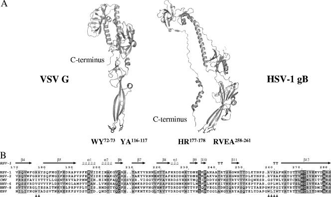
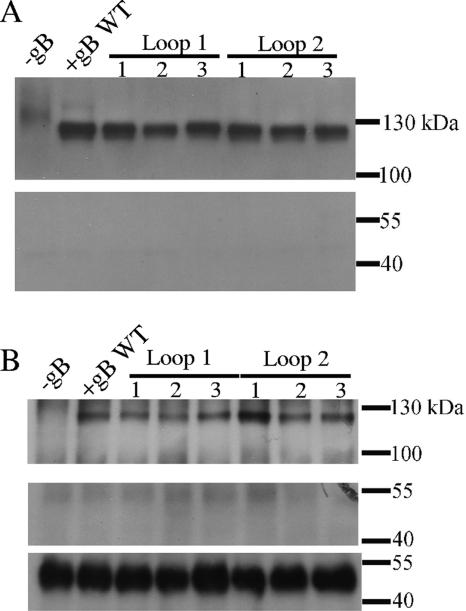
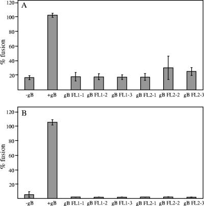
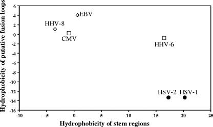
To test the importance of the hydrophobic residues within the putative Epstein-Barr virus (EBV) glycoprotein B (gB) fusion loops in membrane fusion, WY(112-113) and WLIW(193-196) were mutated into alanine, glutamic acid, or the analogous residues from herpes simplex virus type 1 (HSV-1) gB (HR and RVEA). All gB variants exhibited cell surface expression, demonstrating that the substitutions did not perturb gB trafficking. None of six gB variants was, however, capable of mediating fusion with either epithelial or B cells. These data demonstrate that the bulky and hydrophobic EBV loop residues, which differ from the more hydrophilic HSV-1 residues and appear more compatible with membrane insertion, are essential for EBV gB-dependent fusion.


Figures

FIG. 1.
FIG. 1.
(A) Structures of the ectodomains of HSV-1 gB and G protein of VSV in postfusion conformations. Structural homology is notable between HSV-1 gB and VSV G protein, despite the lack of similarity at the protein sequence level. For clarity reasons, only monomers are shown. Residues forming a bipartite fusion peptide in VSV G protein (fusion loop 1, WY72-73; fusion loop 2, YA116-117) are labeled, and their side chains are shown as sticks. Fusion loops in the VSV G protein adopt a hairpin conformation that is typical for the internal fusion peptides of class II fusion proteins and that is compatible with membrane penetration. Residues located in the structurally homologous loops in HSV-1 gB are marked, and their side chains are shown as sticks (fusion loop 1, HR177-178; fusion loop 2, RVEA258-261). The conformations of HSV-1 gB loops do not resemble a hairpin fold and seem to be suboptimal for membrane insertion. The corresponding residues in EBV gB (fusion loop 1, WY112-113; fusion loop 2, WLIW193-196) were mutated in this study to evaluate their importance for the ability of EBV gB to mediate fusion. Both HSV-1 gB and VSV G ectodomains used for crystallization were truncated at the C terminus just before the stem regions. C termini are marked in both structures to indicate the putative location of the stem regions. Protein Data Bank files used for this figure are 2gum and 2cmx. The figure was generated using PyMOL (4). (B) Sequence alignment of gB fragments containing putative fusion loops. In contrast to the highly conserved fusion peptides identified for G protein and class I (5) and class II fusion proteins (1), the fusion loops of different gB proteins are not well conserved. Protein sequences are shown for representative herpesviruses known to infect humans: HSV-1 and HSV-2, cytomegalovirus (CMV) HHV-6, HHV-8, and EBV. Secondary structure elements, extracted from the HSV-1 gB ectodomain X-ray structure (Protein Data Bank file 2gum), are shown on top. Numbering is shown for unprocessed HSV-1 gB. Locations of the residues proposed to form fusion loops in gB are marked with triangles at the bottom of the alignment. Amino acids in the putative fusion loops in HSV-1 and EBV gB are boxed. Amino acids are shaded according to their conservation. The Risler matrix (28) was used to calculate similarity scores. Residues showing strict conservation are shown in dark gray, and residues with a similarity score of 0.7 and higher are shown in light gray. Alignment was generated using the ESPript program (8). Swiss-Prot entry numbers for the sequences shown are (from top to bottom) P10211, P06763, P06473, P36319, P03188, and P88906.
FIG. 2.
FIG. 2.
Surface expression of EBV gB variants. Surface proteins were biotinylated, and neutravidin beads were used to precipitate all biotinylated proteins (A); alternatively, protein G beads loaded with anti-gB antibodies (upper panel) or anti-actin antibodies (middle and lower panels) were used to precipitate gB or actin, respectively (B). In the upper part of panel A, biotinylated gB was detected by Western blotting using anti-gB antibody, while biotinylated actin could not be detected when anti-actin antibody was used instead (lower panel). In panel B avidin conjugated to horseradish peroxidase was used for detection of biotinylated proteins in the upper and middle blots. Biotinylated gB was detected (upper panel), and there was no biotinylated actin in the samples (middle panel). Actin was detected when anti-actin antibody was used for detection, confirming the presence of actin. Samples are labeled as shown in Table 1. WT, wild type.
FIG. 3.
FIG. 3.
Effect of the amino acid substitutions in EBV gB putative fusion loops on epithelial cell fusion (A) and B-cell fusion (B). CHO-K1 cells were transiently transfected with plasmids encoding gB (wild-type or mutant protein), gH/gL, and T7-driven luciferase for epithelial cells fusion or gB (wild-type or mutant protein), gH/gL, gp42, and T7-driven luciferase for B-cell fusion. The CHO-K1 cells were overlaid with the same number of 293T epithelial cells or Daudi B cells expressing T7 polymerase. Fusion was allowed to proceed for 24 h, and luciferase activity was measured to quantify the level of fusion. The first sample lacked gB (gB−) and served as a background control. The second sample (+gB) refers to the positive control (transfected with wild-type gB, gH/gL, and T7-driven luciferase for epithelial cell fusion and gp42 for B-cell fusion). gB mutant proteins are marked according to the type of putative fusion loop (FL) substitution shown in Table 1. Luciferase activity measured for the positive control is set to 100%, and the rest of the measurements are expressed as a percentage of the positive control.
FIG. 4.
FIG. 4.
Negative correlation of hydrophobicities of gB putative fusion loops and stem regions. Total hydrophobicities were calculated using the Kyle and Doolittle scale. Protein sequences and abbreviations used here are the same as shown in Fig. 1B. Residues found in the putative fusion loops and used in calculations are labeled with filled triangles at the bottom in Fig. 1B. The region spanning 50 residues located immediately upstream of the transmembrane anchor (residues 774 to 795) in HSV-1 gB (27) contains two hydrophobic helical regions which, we hypothesized, might serve as stem regions in HSV-1 gB. The borders of homologous segments in gB proteins of other herpesviruses were determined based on the gB sequence alignment (data not shown). The hydrophobicity values calculated for putative fusion loops are plotted as a function of hydrophobicity values obtained for the stem regions. Due to the higher hydrophobicity of stem regions and lower hydrophobicity of the fusion loops, gB of α-herpesviruses HSV-1 and HSV-2 cluster at the opposite side of the plot from the gB of γ-herpesviruses EBV and HHV-8, which have more hydrophobic fusion loops and fewer hydrophobic stem regions.

References

    1. Allison, S. L., J. Schalich, K. Stiasny, C. W. Mandl, and F. X. Heinz. 2001. Mutational evidence for an internal fusion peptide in flavivirus envelope protein E. J. Virol. 75:4268-4275. - PMC - PubMed
    1. Burnette, W. N. 1981. “Western blotting”: electrophoretic transfer of proteins from sodium dodecyl sulfate-polyacrylamide gels to unmodified nitrocellulose and radiographic detection with antibody and radioiodinated protein A. Anal. Biochem. 112:195-203. - PubMed
    1. Daniels, G. M., and S. G. Amara. 1998. Selective labeling of neurotransmitter transporters at the cell surface. Methods Enzymol. 296:307-318. - PubMed
    1. DeLano, W. L. 2002. The PyMOL molecular graphics system. DeLano Scientific, San Carlos, CA.
    1. Dutch, R. E., T. S. Jardetzky, and R. A. Lamb. 2000. Virus membrane fusion proteins: biological machines that undergo a metamorphosis. Biosci. Rep. 20:597-612. - PubMed

Publication types

Substances

LinkOut - more resources