. 2007 Aug;81(16):8821-6.
doi: 10.1128/JVI.00754-07. Epub 2007 Jun 6.

Reversible inhibition of the fusion activity of measles virus F protein by an engineered intersubunit disulfide bridge

Affiliations

Reversible inhibition of the fusion activity of measles virus F protein by an engineered intersubunit disulfide bridge

Jin K Lee et al. J Virol. 2007 Aug.

Abstract

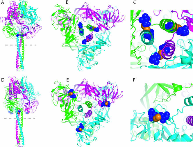
In search of target sites for the development of paramyxovirus inhibitors, we have engineered disulfide bridges to introduce covalent links into the prefusion F protein trimer of measles virus. F-Edm-452C/460C, predicted to bridge head and stalk domains of different F monomers, shows a high degree of proteolytic maturation and surface expression, predominantly as stable, dithiothreitol-sensitive trimers, but no fusion activity. Reduction of disulfide bridges partially restores activity. These findings underscore the importance of reversible intersubunit interactions between the stalk and head domains for F activity. Noncovalent small molecules mimicking this behavior may constitute a potent strategy for preventing paramyxovirus entry.


Figures

FIG. 1.
FIG. 1.
Predicted geometries of disulfide bonds introduced into the prefusion MV F trimer. Homology models of MV F were generated on the basis of the coordinates reported for prefusion PIV5 F and are colored by monomer subunits. (A, B, and C) A disulfide bond between residues 452 and 460 is predicted to establish an intersubunit link between the top of the HR-B prefusion stalk and the base of the globular head domain. Side view of the trimer (A), view from the dotted line in panel A up the HR-B stalk (B), and close-up view of the intersection of stalk and head domain (C). Cysteine side chains of engineered bonds are highlighted in blue; individual sulfur atoms are shown in orange. (D, E, and F) A disulfide bond between residues 307 and 448 is predicted to link two loops in the base of the prefusion head domain of the same monomer. Individual views and coloring of engineered bonds as described for panels A through C.
FIG. 2.
FIG. 2.
An F-Edm-452C/460C double mutant is proteolytically activated and intracellular transport competent but lacks fusion activity. (A) Immunoprecipitation (IP) of F-Edm, F-Edm-452C/460C, and F-Edm-307C/448C from cleared Vero cell lysates obtained 36 h posttransfection. An antiserum directed against residues 127 to 193 in the F HR-A domain was employed. Precipitated material was subsequently subjected to immunostaining using antibodies directed against the cytosolic F tail as previously described (19). Both the F0 precursor and the proteolytically matured F1 fraction of F-Edm and F-Edm-452C/460C are present, while for F-Edm-307C/448C, only the uncleaved F0 precursor is visible. Samples were fractionated under reducing conditions, mock-transfected cells did not express any MV F antigenic material, and immunoblots were decorated with rabbit im- munoglobulin G light-chain-specific horseradish peroxidase conjugate. (B) The F-Edm-452C/460C mutant but not F-Edm-307C/448C is expressed at the cell surface. Surface biotinylation to assess plasma membrane steady-state levels of MV F was carried out as described previously (16). Values above the blot are based on densitometric quantification of signal intensities using a VersaDoc imaging station. Averages of the results of two independent experiments and standard error of the mean values are presented. (C) Quantitative cell-to-cell fusion assays reveal that both F-Edm variants lack fusion activity. Effector Vero cells were infected with MVA-T7 and cotransfected with 3 μg plasmid DNA, each encoding H-Edm or the F construct specified. Target cells harbored a luciferase reporter construct under the control of the T7 promoter, and luciferase activities as an indicator of cell-to-cell fusion activity were assessed 200 min postmixing of both populations. Mock effector cells received only MVA-T7 and H-Edm encoding plasmid. Bar graphs and numbers show averages and standard deviation values of the results of three independent experiments.
FIG. 3.
FIG. 3.
Disruption of F-Edm-452C/460C trimers requires reducing conditions, indicating presence of intersubunit disulfide bonds. (A) Surface immunoprecipitation (IP) of F-Edm, F-Edm-452C/460C, and F-Edm-307C/448C using an antiserum directed against the HR-A domain, followed by gel electrophoresis under reducing (in the presence of 1.5% DTT) and nonreducing (in the absence of DTT) conditions and immunodetection with F-tail-specific antibodies. Under nonreducing conditions, the majority of the F-Edm-452C/460C antigenic material migrates as trimer, while parental F-Edm migrates exclusively as monomer. Reduction of disulfide bonds through DTT treatment results in migration of the majority of F-Edm and F-Edm-452C/460C antigenic material as F1 monomers. Immunoblots were decorated with rabbit immunoglobulin G light-chain-specific horseradish peroxidase conjugate. (B) Fractionation of cell lysates harboring F-Edm-452C/460C on 10 to 25% sucrose gradients, carried out essentially as described previously (14, 18). Prior to loading on gradients, each lysate was treated for 30 min with 1% SDS or 1% SDS and 0.1 M DTT or left untreated (w/o). The gradient fractions were subjected to trichloroacetic acid precipitation, followed by gel electrophoresis under reducing conditions. No redistribution of the F-Edm-452C/460C material to a lower density fraction occurs upon treatment with SDS alone.
FIG. 4.
FIG. 4.
DTT treatment of cells coexpressing H-Edm and F-Edm-452C/460C results in partial reactivation of F-Edm-452C/460C fusion activity. (A) (Top two rows) microphotographs of Vero cells cotransfected with 3 μg plasmid DNA each encoding H-Edm or F-Edm, or encoding H-Edm or F-Edm-452C/460C, or transfected with F-Edm encoding plasmids alone. Thirty hours posttransfection, cells were treated with DTT or left untreated (w/o), followed by an iodoacetamide wash and microscopic assessment of fusion activity 150 min posttreatment. F-Edm-452C/460C-expressing cells formed syncytia only after treatment with DTT. Prior to the time of DTT treatment, H-Edm/F-Edm-expressing cells were kept in the presence of fusion inhibitory peptide (Bachem) to prevent premature syncytia formation. (Bottom row) cells cotransfected with H-Edm and F-Edm-452C/460C as described above but treated with 6.25, 12.5, or 50 mM DTT for 150 min for comparison. The most substantial activation of F-Edm-452C/460C occurs when cells were treated with 12.5 or 25 mM DTT. All microphotographs were taken at a magnification of ×200. (B) Quantification of fusion activity of cells transfected as in panel A using the luciferase reporter assay outlined in Fig. 2C. While treatment with 25 mM DTT reduces fusion activity of F-Edm by approximately 40% compared to untreated (w/o) H-Edm/F-Edm-expressing cells, it restores activity of F-Edm-452C/460C to levels corresponding to 20% of untreated F-Edm. No fusion activity was detected in untreated cells expressing H-Edm and F-Edm-452C/460C. Luciferase activities were normalized for values obtained for untreated, H-Edm/F-Edm-expressing cells. Averages and standard error of the mean values are shown.

Similar articles

Cited by

References

    1. Cathomen, T., H. Y. Naim, and R. Cattaneo. 1998. Measles viruses with altered envelope protein cytoplasmic tails gain cell fusion competence. J. Virol. 72:1224-1234. - PMC - PubMed
    1. Centers for Disease Control and Prevention. 2005. Progress in reducing measles mortality—worldwide, 1999-2003. Morb. Mortal. Wkly. Rep. 54:200-203. - PubMed
    1. Chenna, R., H. Sugawara, T. Koike, R. Lopez, T. J. Gibson, D. G. Higgins, and J. D. Thompson. 2003. Multiple sequence alignment with the Clustal series of programs. Nucleic Acids Res. 31:3497-3500. - PMC - PubMed
    1. Connolly, S. A., G. P. Leser, H. S. Yin, T. S. Jardetzky, and R. A. Lamb. 2006. Refolding of a paramyxovirus F protein from prefusion to postfusion conformations observed by liposome binding and electron microscopy. Proc. Natl. Acad. Sci. USA 103:17903-17908. - PMC - PubMed
    1. Doyle, J., A. Prussia, L. K. White, A. Sun, D. C. Liotta, J. P. Snyder, R. W. Compans, and R. K. Plemper. 2006. Two domains that control prefusion stability and transport competence of the measles virus fusion protein. J. Virol. 80:1524-1536. - PMC - PubMed

Publication types

MeSH terms

LinkOut - more resources