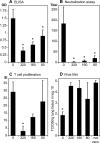
Measles virus-specific CD4 T-cell activity does not correlate with protection against lung infection or viral clearance
- PMID: 17553890
- PMCID: PMC1951373
- DOI: 10.1128/JVI.00160-07
Measles virus-specific CD4 T-cell activity does not correlate with protection against lung infection or viral clearance
Abstract
Acute measles in children can be prevented by immunization with the live attenuated measles vaccine virus. Although immunization is able to induce CD4 and CD8 T cells as well as neutralizing antibodies, only the latter have been correlated with protective immunity. CD8 T cells, however, have been documented to be important in viral clearance in the respiratory tract, whereas CD4 T cells have been shown to be protective in a mouse encephalitis model. In order to investigate the CD4 T-cell response in infection of the respiratory tract, we have defined a T-cell epitope in the hemagglutinin (H) protein for immunization and developed a monoclonal antibody for depletion of CD4 T cells in the cotton rat model. Although the kinetics of CD4 T-cell development correlated with clearance of virus, the depletion of CD4 T cells during the primary infection did not influence viral titers in lung tissue. Immunization with the H epitope induced a CD4 T-cell response but did not protect against infection. Immunization in the presence of maternal antibodies resulted in the development of a CD4 T-cell response which (in the absence of neutralizing antibodies) did not protect against infection. In summary, CD4 T cells do not seem to protect against infection after immunization and do not participate in clearance of virus infection from lung tissue during measles virus infection. We speculate that the major role of CD4 T cells is to control and clear virus infection from other affected organs like the brain.
Figures


Similar articles
-
Sindbis virus-based measles DNA vaccines protect cotton rats against respiratory measles: relevance of antibodies, mucosal and systemic antibody-secreting cells, memory B cells, and Th1-type cytokines as correlates of immunity.J Virol. 2009 Mar;83(6):2789-94. doi: 10.1128/JVI.02191-08. Epub 2009 Jan 7. J Virol. 2009. PMID: 19129445 Free PMC article.
-
Successful mucosal immunization of cotton rats in the presence of measles virus-specific antibodies depends on degree of attenuation of vaccine vector and virus dose.J Gen Virol. 2003 Aug;84(Pt 8):2145-2151. doi: 10.1099/vir.0.19050-0. J Gen Virol. 2003. PMID: 12867646
-
A hemagglutinin-derived peptide-vaccine ignored by virus-neutralizing passive antibodies, protects against murine measles encephalitis.Vaccine. 1999 May 14;17(19):2436-45. doi: 10.1016/s0264-410x(99)00008-0. Vaccine. 1999. PMID: 10392626
-
Neutralizing B cell response in measles.Viral Immunol. 2002;15(3):451-71. doi: 10.1089/088282402760312331. Viral Immunol. 2002. PMID: 12479395 Review.
-
The Immune Response in Measles: Virus Control, Clearance and Protective Immunity.Viruses. 2016 Oct 12;8(10):282. doi: 10.3390/v8100282. Viruses. 2016. PMID: 27754341 Free PMC article. Review.
Cited by
-
Correlates of protection induced by vaccination.Clin Vaccine Immunol. 2010 Jul;17(7):1055-65. doi: 10.1128/CVI.00131-10. Epub 2010 May 12. Clin Vaccine Immunol. 2010. PMID: 20463105 Free PMC article. Review.
-
Sindbis virus-based measles DNA vaccines protect cotton rats against respiratory measles: relevance of antibodies, mucosal and systemic antibody-secreting cells, memory B cells, and Th1-type cytokines as correlates of immunity.J Virol. 2009 Mar;83(6):2789-94. doi: 10.1128/JVI.02191-08. Epub 2009 Jan 7. J Virol. 2009. PMID: 19129445 Free PMC article.
-
Cooperation of Oligodeoxynucleotides and Synthetic Molecules as Enhanced Immune Modulators.Front Nutr. 2019 Aug 27;6:140. doi: 10.3389/fnut.2019.00140. eCollection 2019. Front Nutr. 2019. PMID: 31508424 Free PMC article. Review.
-
Cytokine imbalance after measles virus infection has no correlation with immune suppression.J Virol. 2009 Jul;83(14):7244-51. doi: 10.1128/JVI.00148-09. Epub 2009 May 6. J Virol. 2009. PMID: 19420081 Free PMC article.
-
Synergistic induction of interferon α through TLR-3 and TLR-9 agonists stimulates immune responses against measles virus in neonatal cotton rats.Vaccine. 2014 Jan 3;32(2):265-70. doi: 10.1016/j.vaccine.2013.11.013. Epub 2013 Nov 18. Vaccine. 2014. PMID: 24262312 Free PMC article.
References
-
- Bruton, O. C. 1953. Agammaglobulinemia. Pediatrics 9:722-728. - PubMed
-
- Chen, R. T., L. E. Markowitz, P. Albrecht, J. A. Stewart, L. M. Mofenson, S. R. Preblud, and W. A. Orenstein. 1990. Measles antibody: reevalution of protective titers. J. Infect. Dis. 162:1036-1042. - PubMed
-
- Gans, H., R. DeHovitz, B. Forghani, J. Beeler, Y. Maldonado, and A. M. Arvin. 2003. Measles and mumps vaccination as a model to investigate the developing immune system: passive and active immunity during the first year of life. Vaccine 21:3398-3405. - PubMed
MeSH terms
Substances
LinkOut - more resources
Full Text Sources
Medical
Molecular Biology Databases
Research Materials

