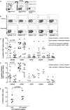
Decreased CXCR3+ CD8 T cells in advanced human immunodeficiency virus infection suggest that a homing defect contributes to cytotoxic T-lymphocyte dysfunction
- PMID: 17553894
- PMCID: PMC1951383
- DOI: 10.1128/JVI.00199-07
Decreased CXCR3+ CD8 T cells in advanced human immunodeficiency virus infection suggest that a homing defect contributes to cytotoxic T-lymphocyte dysfunction
Abstract
To exert their cytotoxic function, cytotoxic T-lymphocytes (CTL) must be recruited into infected lymphoid tissue where the majority of human immunodeficiency virus (HIV) replication occurs. Normally, effector T cells exit lymph nodes (LNs) and home to peripheral sites of infection. How HIV-specific CTL migrate into lymphoid tissue from which they are normally excluded is unknown. We investigated which chemokines and receptors mediate this reverse homing and whether impairment of this homing could contribute to CTL dysfunction as HIV infection progresses. Analysis of CTL chemokine receptor expression in the blood and LNs of untreated HIV-infected individuals with stable, chronic infection or advanced disease demonstrated that LNs were enriched for CXCR3(+) CD8 T cells in all subjects, suggesting a key role for this receptor in CTL homing to infected lymphoid tissue. Compared to subjects with chronic infection, however, subjects with advanced disease had fewer CXCR3(+) CD8 T cells in blood and LNs. CXCR3 expression on bulk and HIV-specific CD8 T cells correlated positively with CD4 count and negatively with viral load. In advanced infection, there was an accumulation of HIV-specific CD8 T cells at the effector memory stage; however, decreased numbers of CXCR3(+) CD8 T cells were seen across all maturation subsets. Plasma CXCL9 and CXCL10 were elevated in both infected groups in comparison to the levels in uninfected controls, whereas lower mRNA levels of CXCR3 ligands and CD8 in LNs were seen in advanced infection. These data suggest that both CXCR3(+) CD8 T cells and LN CXCR3 ligands decrease as HIV infection progresses, resulting in reduced homing of CTL into LNs and contributing to immune dysfunction.
Figures


Similar articles
-
CXC chemokines IP-10 and mig expression and direct migration of pulmonary CD8+/CXCR3+ T cells in the lungs of patients with HIV infection and T-cell alveolitis.Am J Respir Crit Care Med. 2000 Oct;162(4 Pt 1):1466-73. doi: 10.1164/ajrccm.162.4.2003130. Am J Respir Crit Care Med. 2000. PMID: 11029363
-
Selective Expression of CCR10 and CXCR3 by Circulating Human Herpes Simplex Virus-Specific CD8 T Cells.J Virol. 2017 Sep 12;91(19):e00810-17. doi: 10.1128/JVI.00810-17. Print 2017 Oct 1. J Virol. 2017. PMID: 28701399 Free PMC article.
-
Relationship of HIV-1 provirus load, CD8+ CD11+ T cells and HIV-1 envelope-specific cytotoxic T lymphocytes in HIV-infected asymptomatic offients.Immunology. 1994 Sep;83(1):81-5. Immunology. 1994. PMID: 7821971 Free PMC article.
-
CD8 lymphocytes in HIV infection: helpful and harmful.J Clin Lab Immunol. 1997;49(1):15-32. J Clin Lab Immunol. 1997. PMID: 9819670 Review.
-
HIV-specific CD8+ T cells: serial killers condemned to die?Curr HIV Res. 2004 Apr;2(2):153-62. doi: 10.2174/1570162043484960. Curr HIV Res. 2004. PMID: 15078179 Review.
Cited by
-
In vivo Ebola virus infection leads to a strong innate response in circulating immune cells.BMC Genomics. 2016 Sep 5;17(1):707. doi: 10.1186/s12864-016-3060-0. BMC Genomics. 2016. PMID: 27595844 Free PMC article.
-
Low expression of activation marker CD69 and chemokine receptors CCR5 and CXCR3 on memory T cells after 2009 H1N1 influenza A antigen stimulation in vitro following H1N1 vaccination of HIV-infected individuals.Hum Vaccin Immunother. 2015;11(9):2253-65. doi: 10.1080/21645515.2015.1051275. Epub 2015 Jun 19. Hum Vaccin Immunother. 2015. PMID: 26091502 Free PMC article.
-
The colocalization potential of HIV-specific CD8+ and CD4+ T-cells is mediated by integrin β7 but not CCR6 and regulated by retinoic acid.PLoS One. 2012;7(3):e32964. doi: 10.1371/journal.pone.0032964. Epub 2012 Mar 28. PLoS One. 2012. PMID: 22470433 Free PMC article.
-
HIV-1 Nef impairs heterotrimeric G-protein signaling by targeting Gα(i2) for degradation through ubiquitination.J Biol Chem. 2012 Nov 30;287(49):41481-98. doi: 10.1074/jbc.M112.361782. Epub 2012 Oct 15. J Biol Chem. 2012. PMID: 23071112 Free PMC article.
-
Alterations in T and B cell function persist in convalescent COVID-19 patients.Med. 2021 Jun 11;2(6):720-735.e4. doi: 10.1016/j.medj.2021.03.013. Epub 2021 Mar 31. Med. 2021. PMID: 33821250 Free PMC article.
References
-
- Agostini, C., M. Facco, M. Siviero, D. Carollo, S. Galvan, A. M. Cattelan, R. Zambello, L. Trentin, and G. Semenzato. 2000. CXC chemokines IP-10 and Mig expression and direct migration of pulmonary CD8+/CXCR3+ T cells in the lungs of patients with HIV infection and T-cell alveolitis. Am. J. Respir. Crit. Care Med. 162:1466-1473. - PubMed
-
- Appay, V., P. R. Dunbar, M. Callan, P. Klenerman, G. M. Gillespie, L. Papagno, G. S. Ogg, A. King, F. Lechner, C. A. Spina, S. Little, D. V. Havlir, D. D. Richman, N. Gruener, G. Pape, A. Waters, P. Easterbrook, M. Salio, V. Cerundolo, A. J. McMichael, and S. L. Rowland-Jones. 2002. Memory CD8+ T cells vary in differentiation phenotype in different persistent virus infections. Nat. Med. 8:379-385. - PubMed
-
- Aukrust, P., F. Muller, and S. S. Froland. 1998. Circulating levels of RANTES in human immunodeficiency virus type 1 infection: effect of potent antiretroviral therapy. J. Infect. Dis. 177:1091-1096. - PubMed
-
- Brodie, S. J., D. A. Lewinsohn, B. K. Patterson, D. Jiyamapa, J. Krieger, L. Corey, P. D. Greenberg, and S. R. Riddell. 1999. In vivo migration and function of transferred HIV-1-specific cytotoxic T cells. Nat. Med. 5:34-41. - PubMed
Publication types
MeSH terms
Substances
Grants and funding
LinkOut - more resources
Full Text Sources
Medical
Research Materials

