. 2007 Aug;18(8):3081-93.
doi: 10.1091/mbc.e07-02-0172. Epub 2007 Jun 6.

Naked2 acts as a cargo recognition and targeting protein to ensure proper delivery and fusion of TGF-alpha containing exocytic vesicles at the lower lateral membrane of polarized MDCK cells

Affiliations

Naked2 acts as a cargo recognition and targeting protein to ensure proper delivery and fusion of TGF-alpha containing exocytic vesicles at the lower lateral membrane of polarized MDCK cells

Cunxi Li et al. Mol Biol Cell. 2007 Aug.

Abstract

Transforming growth factor-alpha (TGF-alpha) is the major autocrine EGF receptor ligand in vivo. In polarized epithelial cells, proTGF-alpha is synthesized and then delivered to the basolateral cell surface. We previously reported that Naked2 interacts with basolateral sorting determinants in the cytoplasmic tail of a Golgi-processed form of TGF-alpha and that TGF-alpha is not detected at the basolateral surface of Madin-Darby canine kidney (MDCK) cells expressing myristoylation-deficient (G2A) Naked2. By high-resolution microscopy, we now show that wild-type, but not G2A, Naked2-associated vesicles fuse at the plasma membrane. We further demonstrate that Naked2-associated vesicles are delivered to the lower lateral membrane of polarized MDCK cells independent of mu1B adaptin. We identify a basolateral targeting segment within Naked2; residues 1-173 redirect NHERF-1 from the apical cytoplasm to the basolateral membrane, and internal deletion of residues 37-104 results in apical mislocalization of Naked2 and TGF-alpha. Short hairpin RNA knockdown of Naked2 leads to a dramatic reduction in the 16-kDa cell surface isoform of TGF-alpha and increased cytosolic TGF-alpha immunoreactivity. We propose that Naked2 acts as a cargo recognition and targeting (CaRT) protein to ensure proper delivery, tethering, and fusion of TGF-alpha-containing vesicles to a distinct region at the basolateral surface of polarized epithelial cells.


Figures

Figure 1.
Figure 1.
Naked2-associated vesicle fusion events. (A) Domains of human Naked2. Glycine, the second residue, undergoes myristoylation. Naked homologue region 1 (NHR1) consists of residues 107-175; NHR2 includes residues 236-265; TGFα tail-binding domain (TTB) was mapped to residues 300-385 (Li et al., 2004), and H16 represents the last 22 residues that contain 16 histidines. (B) Live cell microscopy of a typical homotypic vesicle–vesicle fusion event. Bar, 1 μm. (C) For vesicles 1, 2, and 3, the integrated intensities (area X gray scale) were quantified by Metamorph software. (D) Time-lapse images were captured by total internal reflection fluorescent microscopy (TIR-FM) to visualize vesicle fusion at the plasma membrane. MDCK cells stably transfected with wild-type Naked2-EGFP were seeded onto MatTek dishes for 24 h, and then TIR-FM was performed. The microscope stage was maintained at 34°C. Scanning time was 200 ms for each 512 × 512 pixel image at 500-ms intervals. The dashed line indicates a region where a vesicle fusion event occurs. Quantification of fluorescent intensity is shown under each selected region. Bars, 2 μm.
Figure 2.
Figure 2.
Live cell microscopy of Naked2-associated vesicles in polarized MDCK cells. (A) MDCK cells stably transfected with Naked2-EGFP were plated on the undersurface of Transwell filters, and experiments were performed on day 4 when electrical resistance exceeded 200 Ω/cm2. Time-lapse images were taken from six sequential planes from the midpoint to the base of the cell at 2.5-s intervals as indicated in the top right corner of B. The green arrow in A indicates a representative vesicle visualized at the 5th plane. Fluorescent intensity of the vesicle diminishes over time in this plane. (B) The relative fluorescent intensity of the selected vesicle in A was manually traced, tracked, and quantified in six Z-sections at 20 time points over the last 60 s of the time-lapse movie. Bar, 10 μm.
Figure 3.
Figure 3.
Tannic acid prevents cell surface fusion of Naked2-associated vesicles at the lower portion of the lateral plasma membrane of polarized MDCK cells. (A) MDCK cells stably transfected with Naked2-EGFP cDNA were cultured on Transwell filters for 4 d and then placed at 40°C for 4 h; this “heat bleaching” resulted in disappearance of GFP fluorescence. (B) After this 40°C heat bleaching, cells were then placed at 10°C for 10 min with 0.5% tannic acid added selectively to the basolateral medium, and then cells were restored to 37°C for 15 min in the continued presence of tannic acid. At this time, Naked2 fluorescence appeared at the lower lateral corner. (C) When cells were restored to 37°C for 2 h after 40°C heat bleaching in the absence of tannic acid, Naked2 fluorescence was found along the entire lateral membrane. F-actin was stained with rhodamine-phalloidin (red) to outline the cell borders. Mean fluorescent intensity ± SD from at least three separate experiments is shown under the representative image. Bar, 10 μm.
Figure 4.
Figure 4.
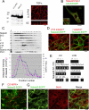
Basolateral delivery of Naked2-associated vesicles does not require μ1B. (A) Endogenous Naked2 was detected by Western blotting of whole cell lysates from these LLC-PK1 cells and Caco-2 cells (a human colon cancer cell line) using R44, a previously described rabbit polyclonal antibody to human Naked2 (Li et al., 2004). HA-tagged TGFα was stably transfected into LLC-PK1 cells. TGFα staining decorates the basolateral compartment of these μ1B-deficient LLC-PK1 cells. (B) Transiently transfected Halo-tagged Naked2 did not colocalize with LDL receptor (LDLR) in MDCK cells that had been stably transfected with LDLR-EGFP. (C) Top, Western blotting of Naked2, Dishevelled (DVL1), and endogenous adaptor proteins from fractions isolated from 10 to 40% iodoxinal gradient centrifugation from MDCK cells stably overexpressing Naked2-EGFP and TGFα. Bottom, levels of TGFα by RIA in the different fractions correspond to those enriched for Naked2 by Western blotting. (D) Lack of colocalization of β1/2 adaptin and γ adaptin with Naked2 GFP fluorescence. Antibody to β adaptin (10A) recognizes both β1 and 2 adaptin. mAb 100.3 is specific for γ adaptin. (E) Yeast two-hybrid analysis between Naked2 N1-173 and different AP subunits under histidine minus growth conditions. This Naked2 fragment did not interact directly with AP-1-4 components. Naked2-EGFP did not colocalize with CD-MPR (F), which is sorted by GGA (Puertollano et al., 2001) or Sec6 (G). Bars, 10 μm.
Figure 5.
Figure 5.
Mapping of Naked2 basolateral targeting domain. (A) Fragments of human Naked2 were fused to human Na+/H+ exchanger regulatory factor (NHERF)-1 and dsRed2. These chimeras were transfected stably into MDCK cells. (B) Relative expression of these chimeras by Western blotting. (C) XY and XZ images of polarized MDCK cells expressing different Naked2 and NHERF-1 chimeras. XY images of polarized MDCK cells were obtained at the apex of the cells near the tight junctions (using ZO-1, green, as shown in panels 4 and 5). FR (NHERF-1 fused to dsRed2) showed diffuse staining in the apical cytoplasm of polarized MDCK cells; Naked2 residues 1-265 fused with FR (N1-265-FR) redirected FR to the basolateral plasma membrane like wild-type Naked2. Naked2 residues 1-173 fused with FR (N1-173-FR) also decorated the basolateral plasma membrane. Fusion of N-terminal 1-104 amino acids of Naked2 to FR (N1-104-FR) exhibited strong staining at the apical and basolateral plasma membrane. Naked2 residues 1-36 fused with FR (N1-36-FR) exhibited predominant staining of the apical compartment (cytoplasm and plasma membrane) with weaker staining along the basolateral plasma membrane. Bar, 10 μm.
Figure 6.
Figure 6.
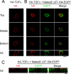
Loss of basolateral selectivity for Naked2 and TGFα with deletion of Naked2 residues 37-104. (A) Normal basolateral membrane localization of HA-tagged TGFα (red) in transiently transfected, polarized MDCK cells. (B) TGFα redistributed apically and partially colocalized with Naked2 Δ37-104-EGFP when MDCK cells were transiently cotransfected with Naked2 Δ37-104-EGFP mutant and HA-tagged TGFα. (C) TGFα was detected at the apical cell surface when HA antibody was added at 4°C to the apical compartment of nonpermeabilized live cells stably cotransfected with HA ectodomain–tagged TGFα and Naked2 Δ37-104-EGFP mutant cDNA constructs. Naked2 fluorescence colocalized with TGFα immunoreactivity at the apical surface. Bars, 10 μm.
Figure 7.
Figure 7.
shRNA knockdown of endogenous Naked2 results in increased cytosolic TGFα immunoreactivity with a dramatic reduction in cell surface TGFα. HaCat cells were stably transfected with an HA-tagged–TGFα cDNA; this clone was designated HaCat23. (A) Retroviral-mediated delivery of Naked2 shRNA to HaCat23 cells reduced endogenous Naked2 levels by 60% compared with vector control and parental cells. (B) There was a significant increase of cytosolic TGFα immunoreactivity in Naked2 shRNA-treated cells, as quantified by reconstructed 3D images of 10 fields from confocal micrographs of whole cells; mean intensity ± SD. Student's t test, p < 0.01. Bar, 10 μm. (C) Western blot of immunoprecipitated TGFα showed a marked reduction in the 16-kDa cell surface isoform and a correspondingly increase of Golgi-processed isoform (36 kDa).

Similar articles

Cited by

References

    1. Apodaca G., Bomsel M., Lindstedt R., Engel J., Frank D., Mostov K. E., Wiener-Kronish J. Characterization of Pseudomonas aeruginosa-induced MDCK cell injury: glycosylation-defective host cells are resistant to bacterial killing. Infect. Immun. 1995;63:1541–1551. - PMC - PubMed
    1. Bonifacino J. S., Glick B. S. The mechanisms of vesicle budding and fusion. Cell. 2004;116:153–166. - PubMed
    1. Bonifacino J. S., Traub L. M. Signals for sorting of transmembrane proteins to endosomes and lysosomes. Annu. Rev. Biochem. 2003;72:395–447. - PubMed
    1. Briley G. P., Hissong M. A., Chiu M. L., Lee D. C. The carboxyl-terminal valine residues of proTGF alpha are required for its efficient maturation and intracellular routing. Mol. Biol. Cell. 1997;8:1619–1631. - PMC - PubMed
    1. Dempsey P. J., Coffey R. J. Basolateral targeting and efficient consumption of transforming growth factor-alpha when expressed in Madin-Darby canine kidney cells. J. Biol. Chem. 1994;269:16878–16889. - PubMed

Publication types

MeSH terms