. 2007 Aug;18(8):3214-23.
doi: 10.1091/mbc.e06-12-1154. Epub 2007 Jun 6.

E-cadherin adhesion activates c-Src signaling at cell-cell contacts

Affiliations

E-cadherin adhesion activates c-Src signaling at cell-cell contacts

Robert W McLachlan et al. Mol Biol Cell. 2007 Aug.

Abstract

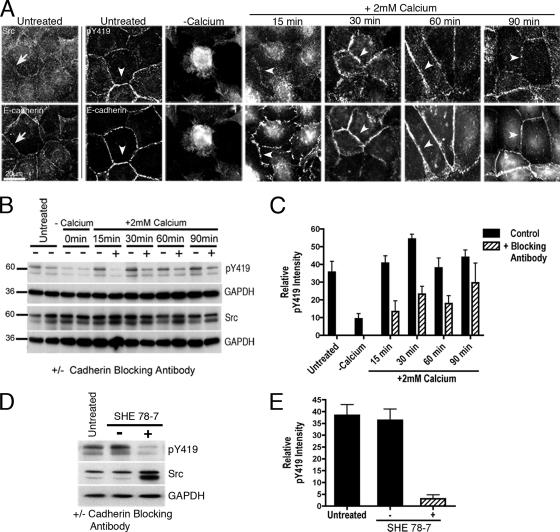
Cadherin-based cell-cell contacts are prominent sites for phosphotyrosine signaling, being enriched in tyrosine-phosphorylated proteins and tyrosine kinases and phosphatases. The functional interplay between cadherin adhesion and tyrosine kinase signaling, however, is complex and incompletely understood. In this report we tested the hypothesis that cadherin adhesion activates c-Src signaling and sought to assess its impact on cadherin function. We identified c-Src as part of a cadherin-activated cell signaling pathway that is stimulated by ligation of the adhesion receptor. However, c-Src has a biphasic impact on cadherin function, exerting a positive supportive role at lower signal strengths, but inhibiting function at high signal strengths. Inhibiting c-Src under circumstances when it is activated by cadherin adhesion decreased several measures of cadherin function. This suggests that the cadherin-activated c-Src signaling pathway serves positively to support cadherin function. Finally, our data implicate PI3-kinase signaling as a target for cadherin-activated c-Src signaling that contributes to its positive impact on cadherin function. We conclude that E-cadherin signaling is an important activator of c-Src at cell-cell contacts, providing a key input into a signaling pathway where quantitative changes in signal strength may result in qualitative differences in functional outcome.


Figures

Figure 1.
Figure 1.
E-cadherin is required for cell–cell contact to activate c-Src. MCF-7 monolayers were studied at confluence (untreated), immediately after cell–cell contacts were broken by chelation of extracellular calcium with 4 mM EDTA (−Calcium), or 15–90 min after replenishing extracellular calcium to allow contacts to reassemble (+2 mM Calcium). (A) Active c-Src is present at established and reassembling cell–cell contacts. Fixed cells were immunostained for E-cadherin and either c-Src (Src, arrows) or activated c-Src (pY419, arrowheads). (B and C) E-cadherin activity is necessary for stimulation of c-Src as cells reassemble contacts with one another. Cells were allowed to reassemble contacts after chelation of extracellular calcium (+2 mM Calcium) either in the presence (+) or absence (−) of an E-cadherin function-blocking antibody (SHE 78-7). Western blots from cell lysates were probed for c-Src, pY419 c-Src, and GAPDH as a loading control; a representative blot is shown in B. pY419 c-Src levels were quantified by densitometry and expressed relative to total c-Src levels (C). Data are means ± SEM (n = 3; p < 0.0001 for cells treated with SHE78-7 for 15, 30 and 60 min). (D and E) E-cadherin is necessary to maintain active c-Src at established cell–cell contacts. Confluent MCF-7 monolayers treated with a cadherin function-blocking antibody (+; SHE 78-7), or with a control Mouse IgG antibody (−), were compared with untreated established monolayers (untreated). Cells were incubated with antibodies for 5 min and then lysed, and Western blots were probed for pY419 c-Src, c-Src, and GAPDH (D). pY419 c-Src levels expressed relative to total c-Src levels (E). Data are means ± SEM (n = 3; p < 0.0001 for SHE 78-7-treated cells compared with control Ab cells).
Figure 2.
Figure 2.
c-Src is activated by E-cadherin homophilic ligation. (A and B) Isolated cultures of MCF-7 cells were incubated for 15–60 min with latex beads coated with either hE/Fc or PLL. These samples were compared with control cell cultures not exposed to beads (−Beads) or control cultures incubated with beads that had been blocked but not coated with any adhesive ligand (+Beads). Western blots from the samples were probed for activated c-Src (pY419 c-Src), total c-Src, and GAPDH (loading control). A representative blot is shown in A. (B) pY419 c-Src levels were measured by densitometry and expressed relative to total c-Src levels. Data are means ± SEM (n = 3; p < 0.0001 at each time point comparing hE/Fc-beads and PLL beads). (C and D) CHO cells expressing wild-type E-cadherin (hE), hE-Cad-764AAA (764), or hE-Cad-PYD-6MT (PYD) were incubated for 15 min with latex beads coated with hE/Fc. In control experiments hE-CHO cells were also incubated with poly-l-lysine–coated beads (PLL). Western blots from cell lysates were probed for pY419 c-Src, c-Src and GAPDH (C). (D) pY419 c-Src levels quantified by densitometry were expressed relative to total c-Src levels. Data are means ± SEM (n = 3).
Figure 3.
Figure 3.
c-Src signaling is required for the integrity of E-cadherin–based cell–cell contacts. (A) MCF-7 cell monolayers transiently expressing dominant-negative (DN) c-Src-251-GFP or DN c-Src-mf (K295M Y527F) were compared with control cells transfected with pEGFP-N1 alone. After fixation, cells were immunostained for E-cadherin and GFP (Tag). Expression of dominant-negative c-Src mutants reduced the intensity of E-cadherin staining found at contacts between two transfected cells (arrows) compared with contacts between either pEGFP-N1 transfected cells or between untransfected cells (arrowheads). (B) Dominant-negative c-Src expression reduces E-cadherin accumulation at epithelial cell–cell contacts. The intensity of E-cadherin staining at transfected cell–cell contacts was measured and expressed as a ratio of the fluorescence intensity at untransfected cell–cell contacts. Data are means ± SEM (n = 30; p < 0.0001 for DN mutants). (C) Inhibition of c-Src with PP2 significantly affects the integrity of epithelial cell–cell contacts. MCF-7 monolayers treated with PP2 (5–10 μM) or PP3 (10 μM) were compared with untreated monolayers. The fluorescence intensity of E-cadherin staining at cell–cell contacts was quantified (Cadherin Intensity). Data are means ± SEM (n = 50; p < 0.0001 for 5–10 μM PP2). (D) Inhibition of c-Src does not affect the total or surface expression of E-cadherin. MCF-7 monolayers trypsinized in the presence (+Ca2+) or absence of extracellular calcium (−Ca2+) were compared with untreated cell monolayers (WCL). Western blots from cell lysates were probed for E-cadherin and β-tubulin as a loading control. (E) The stoichiometry of the cadherin–catenin complex is not significantly affected by c-Src inhibition. Untreated MCF-7 cell monolayers were compared with monolayers treated with either 10 μM PP2 or 10 μM PP3. E-cadherin immunoprecipitates from cell lysates were separated by SDS-PAGE and probed for E-cadherin, β-catenin, α-catenin, and p120.
Figure 4.
Figure 4.
c-Src is required for E-cadherin adhesive interactions. (A) Src signaling is necessary for E-cadherin adhesion. Adhesion of cells to hE/Fc-coated substrata was assessed using a laminar flow assay that measures the ability of cells to resist detachment by laminar flow (Flow rate). CHO cells stably expressing human E-cadherin (hE-CHO cells) were studied after pre-treatment with either PP2 (10 μM) or PP3 (10 μM). Negative controls were parental CHO cells that lack E-cadherin (CHO φ) and hE-CHO cells incubated with uncoated capillary tubes (No hE/Fc). PP2 significantly reduced the adhesiveness of cells compared with PP3-treated cells. Data are means ± SEM (n = 3; p < 0.0001). (B) c-Src is required for cells to spread on cadherin-coated substrata. hE-CHO cells were allowed to adhere and spread on hE/Fc-coated substrata. Cells transiently transfected with dominant-negative (DN) c-Src-251 or DN c-Src-mf (K295M Y527F) were compared with cells expressing pEGFP-N1 alone (GFP) or with untransfected cells. The ability of the cells to form and extend cadherin-based adhesions was quantified by measuring cell surface area. Representative images are shown in insets. Data are means ± SEM (n = 35; p < 0.0001 for DN mutants compared with untransfected controls). (C) PP2 prevents cells from spreading on cadherin-coated substrata. hE-CHO cells incubated with either PP2 (10 μM) or PP3 (10 μM) were allowed to adhere to hE/Fc-coated substrata, and spreading was assessed by measuring cell surface area. Representative images are shown in insets. Withdrawal of drug for 1 h was sufficient to restore spreading in PP2-treated cells (Drug + Washout). Data are means ± SEM (n = 35; p < 0.0001 for PP2 vs. PP3 and p = 0.8 for PP2 + Washout vs. PP3 + Washout).
Figure 5.
Figure 5.
Constitutively active c-Src disrupts E-cadherin function. (A) c-SrcY527F-GFP expression perturbs the morphology of E-cadherin–based cell–cell contacts. MCF-7 cells expressing c-SrcY527F-GFP or EGFP were allowed to form cellular contacts. After fixation, cells were immunostained for E-cadherin and GFP (Tag). In comparison to untransfected or pEGFP-N1 cell–cell contacts (arrowheads), c-SrcY527F-GFP–expressing cells failed to form E-cadherin–based cell–cell contacts (arrows). (B) Constitutively active c-Src prevents the formation and extension of E-cadherin–based contacts. hE-CHO cells expressing c-SrcY527F-GFP or EGFP were allowed to adhere to hE/Fc-coated substrata, and their ability to spread was assessed by measuring the surface area of the cells. Representative images are shown in insets. Data are means ± SEM (n = 30; p < 0.0001).
Figure 6.
Figure 6.
Increasing CA c-Src levels have a bimodal effect on cadherin-based cell spreading. Serum-starved hE-CHO cells transiently transfected with c-Src Y525F-GFP were allowed to adhere to hE/Fc-coated substrata for 90 min. Cell spreading (pixels) was then compared with expression of c-Src Y525F-GFP, assessed by fluorescence intensity of GFP (arbitrary units, au). The curve represents average cell surface area as a function of GFP intensity. Representative images of cells expressing low, medium, or high levels of c-Src Y525F-GFP are shown in insets.
Figure 7.
Figure 7.
c-Src signaling is necessary for E-cadherin to activate PI3-kinase signaling. Isolated MCF-7 cell cultures were incubated for 30–90 min with either hE/Fc coated latex beads (hE/Fc) or PLL-coated beads (PLL). Cells exposed to cadherin-coated beads were incubated with either PP2 (10 μM) or PP3 (10 μM). At each time point, cell lysates were subjected to subcellular fractionation and the membrane-enriched (P100) fraction was probed for Akt, pAkt, and transferrin receptor (TfR) by Western analysis. Additionally, total lysates were probed for Akt and GAPDH as a loading control. A representative of three independent experiments is shown in A. (B) Recruitment of Akt to membranes was quantitated relative to total cellular levels of Akt in the cells. (C) pAkt levels in membranes were quantitated relative to TfR levels in membranes. Data are means ± SEM (n = 3). PP2 reduced the amounts of both Akt (A and B) and pAkt (C) found in membranes in response to cadherin homophilic ligation.
Figure 8.
Figure 8.
PI3-kinase signaling can restore E-cadherin function in c-Src inhibited cells. (A and B) Expression of the constitutively active (CA) PI3-kinase (p110-CAAX) rescues the integrity of c-Src-inhibited cadherin-based contacts. p110-CAAX was transiently expressed in untreated, PP3- and PP2-treated MCF-7 monolayers. After fixation, the monolayers were probed for E-cadherin and anti-myc (Tag) to identify p110-CAAX–expressing cells (A). E-cadherin fluorescence intensity at contacts between untransfected cells (U-U) and cells expressing p110-CAAX (arrowheads, T-T) was quantitated by digital image analysis (B). Data are means ± SEM (n = 30). (C and D) Constitutively active p110-CAAX restores the ability of PP2-treated cells to spread on hE/Fc. Cells transiently transfected with either p110-CAAX or GFP alone were incubated with either PP2 (10 μM) or PP3 (10 μM) and allowed to adhere to hE/Fc-coated substrata. Cells were then stained for either F-actin (Actin) or GFP/Myc (Tag) (C). Cell spreading was quantitated by measuring the surface area of adherent cells (D). Note that expression of p110-CAAX restored the ability of PP2-treated cells to spread on hE/Fc to levels comparable to negative controls (Untreated, GFP) as well as after PP2 was withdrawn (PP2+Washout). Data are means ± SEM (n = 30).

Similar articles

Cited by

References

    1. Avizienyte E., Wyke A. W., Jones R. J., McLean G. W., Westhoff M. A., Brunton V. G., Frame M. C. Src-induced de-regulation of E-cadherin in colon cancer cells requires integrin signalling. Nat. Cell Biol. 2002;4:632–638. - PubMed
    1. Behrens J., Vakaet L., Friis R., Winterhager E., Van Roy F., Mareel M. M., Birchmeier W. Loss of epithelial differentiation and gain of invasiveness correlates with tyrosine phosphorylation of the E-cadherin/beta-catenin complex in cells transformed with a temperature-sensitive v-SRC gene. J. Cell Biol. 1993;120:757–766. - PMC - PubMed
    1. Calautti E., Cabodi S., Stein P. L., Hatzfeld M., Kedersha N., Paolo Dotto G. Tyrosine phosphorylation and src family kinases control keratinocyte cell-cell adhesion. J. Cell Biol. 1998;141:1449–1465. - PMC - PubMed
    1. Calautti E., Grossi M., Mammucari C., Aoyama Y., Pirro M., Ono Y., Li J., Dotto G. P. Fyn tyrosine kinase is a downstream mediator of Rho/PRK2 function in keratinocyte cell-cell adhesion. J. Cell Biol. 2002;156:137–148. - PMC - PubMed
    1. Cantrell D. A. Phosphoinositide 3-kinase signalling pathways. J. Cell Sci. 2001;114:1439–1445. - PubMed

Publication types

Substances

LinkOut - more resources