. 2007 Aug;18(8):3105-18.
doi: 10.1091/mbc.e05-11-1027. Epub 2007 Jun 6.

Parkin-mediated monoubiquitination of the PDZ protein PICK1 regulates the activity of acid-sensing ion channels

Affiliations

Parkin-mediated monoubiquitination of the PDZ protein PICK1 regulates the activity of acid-sensing ion channels

Monica Joch et al. Mol Biol Cell. 2007 Aug.

Abstract

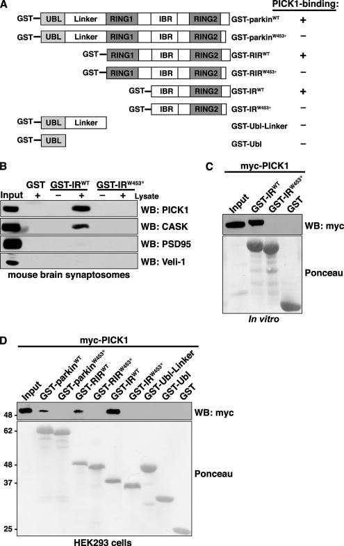
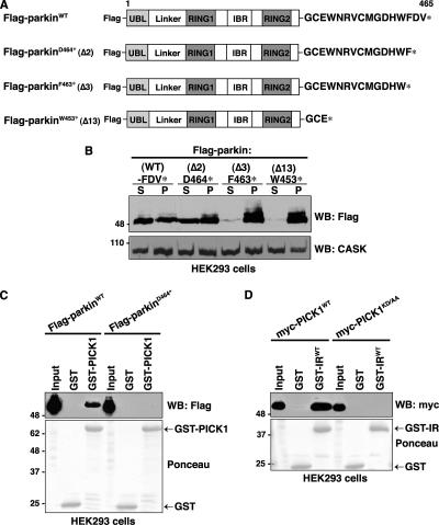
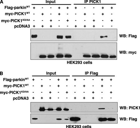
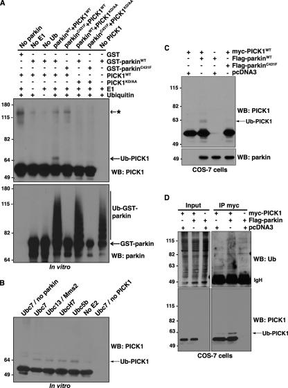
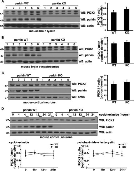
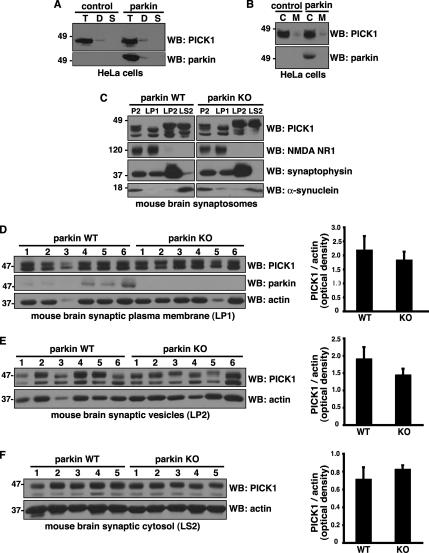
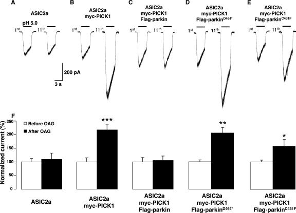
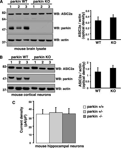
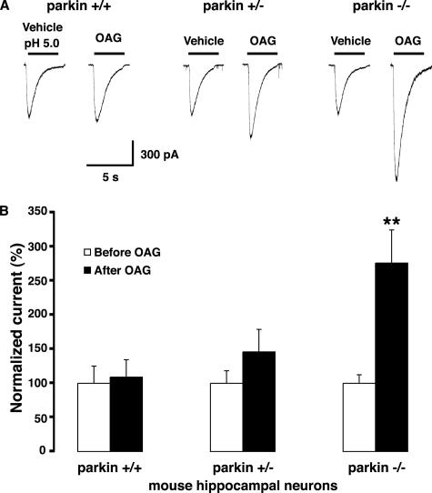
Mutations in the parkin gene result in an autosomal recessive juvenile-onset form of Parkinson's disease. As an E3 ubiquitin-ligase, parkin promotes the attachment of ubiquitin onto specific substrate proteins. Defects in the ubiquitination of parkin substrates are therefore believed to lead to neurodegeneration in Parkinson's disease. Here, we identify the PSD-95/Discs-large/Zona Occludens-1 (PDZ) protein PICK1 as a novel parkin substrate. We find that parkin binds PICK1 via a PDZ-mediated interaction, which predominantly promotes PICK1 monoubiquitination rather than polyubiquitination. Consistent with monoubiquitination and recent work implicating parkin in proteasome-independent pathways, parkin does not promote PICK1 degradation. However, parkin regulates the effects of PICK1 on one of its other PDZ partners, the acid-sensing ion channel (ASIC). Overexpression of wild-type, but not PDZ binding- or E3 ubiquitin-ligase-defective parkin abolishes the previously described, protein kinase C-induced, PICK1-dependent potentiation of ASIC2a currents in non-neuronal cells. Conversely, the loss of parkin in hippocampal neurons from parkin knockout mice unmasks prominent potentiation of native ASIC currents, which is normally suppressed by endogenous parkin in wild-type neurons. Given that ASIC channels contribute to excitotoxicity, our work provides a mechanism explaining how defects in parkin-mediated PICK1 monoubiquitination could enhance ASIC activity and thereby promote neurodegeneration in Parkinson's disease.


Figures

Figure 1.
Figure 1.
PDZ-dependent, direct interaction between parkin and PICK1. (A) GST-parkin fusion constructs are shown with the following functional domains: UBL, ubiquitin-like domain; RING, RING finger motif; IBR, in-between-RINGs motif. Constructs containing the parkin C-terminus encode both the wild-type and truncated PD-linked mutant, W453*. The ability of each GST-parkin construct to interact with wild-type PICK1 is indicated in the right column. (B) The C-terminus of parkin pulls down endogenous PICK1 from mouse brain synaptosomes. Solubilized synaptosomes were incubated with wild-type and mutant parkin C-terminal fusion proteins, GST-IRWT and GST-IRW453*. Binding was assayed by SDS-PAGE and Western blotting with antibodies against candidate PDZ proteins. As shown previously, CASK as well as the PDZ protein PICK1 associate specifically with the wild-type parkin construct and not with the truncated construct or GST alone. (C) PICK1 directly binds the extreme C-terminal residues of parkin in pulldown assays. In vitro–translated myc-PICK1 is selectively pulled down by the wild-type parkin carboxyl-terminal construct, GST-IRWT, and not by the PDZ-binding site mutant, GST-IRW453*, or by GST alone. (D) Domain mapping of interaction of PICK1 with parkin. Myc-PICK1 was transiently expressed in HEK293 cells and pulled down with the indicated GST-parkin fusion proteins or by GST alone. Western blots show that myc-PICK1 is only retained by fusion constructs containing the extreme C-terminal residues of parkin. The ponceau indicates comparable levels of fusion proteins were used in each binding assay.
Figure 2.
Figure 2.
Identification of critical residues involved in the parkin-PICK1 PDZ-mediated interaction. (A) Schematic representation of Flag-parkin constructs. (B) C-terminal truncations exceeding two amino acids render parkin insoluble. HEK293 cells were transiently transfected with the indicated wild-type and C-terminal deletion constructs of parkin. Cells were harvested, lysed in detergent buffer (0.5% Triton X-100 in TBS), and fractionated by centrifugation, and parkin present in the detergent-soluble (S) and -insoluble (P) fractions was analyzed by Western blotting with anti-Flag antibodies. Deletion of the last two residues of parkin (D464*) does not affect parkin solubility compared with the wild-type protein. However, deletion of the final 3 (F463*) or 13 (W453*) amino acids of parkin renders the protein insoluble to mild detergent. Endogenous CASK levels are shown in the bottom panel as a loading control. (C) Deleting the final two amino acids of the PDZ-binding motif (Flag-parkinD464*) of parkin eliminates binding with PICK1 in vitro. Extracts from HEK293 cells transiently transfected with wild-type or PDZ mutant Flag-parkin were incubated with either GST-PICK1 or GST alone. Binding was assayed by SDS-PAGE and Western blotting with the indicated antibodies. (D) Mutation of the PDZ domain of PICK1 disrupts the interaction with parkin. Extracts from HEK293 cells transiently transfected with wild-type or PDZ mutant myc-PICK1 (myc-PICK1KD/AA) were incubated with the indicated GST fusion proteins. Binding was assayed by SDS-PAGE and Western blotting with the indicated antibodies.
Figure 3.
Figure 3.
Parkin and PICK1 associate in cells via a PDZ-mediated interaction. HEK293 cells were transfected with expression vectors encoding Flag-parkin and wild-type myc-PICK1 or myc-PICK1 PDZ mutant K27A,D28A (KD/AA). Extracts were prepared from transfected cells and were immunoprecipitated with (A) anti-PICK1 or (B) anti-Flag antibodies. After separation of immunoprecipitates by SDS-PAGE, Western blotting was performed with the indicated antibodies. In both A and B, parkin associates only with wild-type PICK1. Mutations in the carboxylate-binding loop of PICK1 (KD/AA) inhibit these interactions.
Figure 4.
Figure 4.
Parkin promotes PDZ-dependent PICK1 monoubiquitination. (A) In vitro ubiquitination assays were performed using purified recombinant E1s, E2 (Ubc7), GST-parkin (wild-type or C431F E3 Ub-ligase inactive mutant), PICK1 (wild-type or KD/AA PDZ-mutant), Ub, and ATP. Reagents were combined as indicated at 37°C for 1 h. Reactions were immunoblotted to detect Ub-modified PICK1 species. Monoubiquitination of PICK1 by parkin is dependent on both an intact PDZ domain in PICK1 and functional E3 Ub-ligase activity of parkin. Asterisk (*) indicates nonspecific band, present with or without functional parkin. (B) Multiple E2 enzymes can support parkin-mediated PICK1-monoubiquitination. Ubiquitination reactions were carried out as above with wild-type GST-parkin and PICK1 in the presence of the indicated E2s. (C and D) Parkin enhances the monoubiquitination of PICK1 in cells. (C) Extracts were prepared from COS-7 cells transfected with expression vectors encoding myc-PICK1 and wild-type Flag-parkin or the E3 Ub-ligase inactive parkin mutant Flag-parkinC431F. Proteins were separated by SDS-PAGE, and Western blotting was performed with the indicated antibodies. (D) COS-7 cells were transfected with expression vectors encoding myc-PICK1 and wild-type Flag-parkin. Cells were preincubated with proteasome inhibitor (2 μM lactacystin) for 30 min to allow ubiquitinated species to accumulate. Cells were then lysed in RIPA buffer containing NEM, and soluble proteins were immunoprecipitated with anti-myc antibody. After separation of immunoprecipitates by SDS-PAGE, immunoblotting was used to detect ubiquitinated PICK1.
Figure 5.
Figure 5.
Parkin does not promote PICK1 degradation by the proteasome. (A–C) Steady state endogenous PICK1 levels in mouse whole brain lysates (A), synaptosomes (B), and cortical neurons (C) from parkin wild-type and knockout mice. Equal amounts of protein were loaded from the indicated number of mice and immunoblotted with the antibodies shown. Densitometric analyses of endogenous PICK1 band intensities, normalized to actin, used as a loading control, are presented (right) as mean ± SEM. Student's t test reveals no significant differences between parkin wild-type and knockout samples. (D) Degradation rate of endogenous PICK1 in cortical neurons cultured from parkin wild-type and knockout mice. New protein synthesis was blocked with cycloheximide (40 μg/ml). In parallel cultures, the proteasomal inhibitor lactacystin (2 μM) was applied in addition to cycloheximide. Cells were harvested at the indicated time points after treatment, and protein levels were assessed by immunoblotting with the indicated antibodies. Densitometric analyses of PICK1 band intensities, normalized to actin, are presented (below) as mean ± SEM of five experiments. ANOVA reveals no significant differences in endogenous PICK1 turnover between parkin wild-type and knockout samples regardless of lactacystin treatment.
Figure 6.
Figure 6.
Parkin does not affect PICK1 subcellular localization. (A and B) Subcellular distribution of endogenous PICK1 in HeLa cells stably expressing Flag-parkin or empty vector control. (A) Cells were sequentially extracted with 0.5% Triton X-100 (T), 1% deoxycholic acid (D), and 2% SDS (S). Equal volumes were loaded and immunoblotted with the indicated antibodies. Both PICK1 and parkin were predominantly found in the Triton X-100–soluble fraction with a small amount in the Triton X-100–resistant, deoxycholic acid–soluble fraction. (B) Cells were lysed mechanically without detergent, and the cytosolic (C) and membrane fractions (M) were separated by centrifugation. Similar amounts of protein (volume ratio of 4:1 cytosol:membrane fraction) were loaded and immunoblotted with the indicated antibodies. Both PICK1 and parkin were predominantly found in the cytosolic fraction. Importantly, parkin expression did not change endogenous PICK1 distribution using either the detergent (A) or mechanical (B) fractionation procedure. (C) Distribution of PICK1 in subsynaptic fractions prepared from parkin wild-type and knockout mouse brain synaptosomes (P2). Immunoblotting showed a similar distribution pattern of PICK1 in parkin wild-type and knockout fractions. Antibodies against NMDA NR1, synaptophysin, and α-synuclein were used as markers of synaptic plasma membrane (LP1), synaptic vesicle (LP2), and synaptic cytosolic (LS2) fractions, respectively. (D–F) Quantification of PICK1 levels in synaptic plasma membrane (D), synaptic vesicle (E), and synaptic cytosol (F) from parkin wild-type and knockout mice. Equal amounts of protein were loaded from the indicated number of mice and immunoblotted with the antibodies shown. Densitometric analyses of endogenous PICK1 band intensities, normalized to actin, used as a loading control, are presented (right) as mean ± SEM. Student's t test reveals no significant differences between parkin wild-type and knockout samples.
Figure 7.
Figure 7.
Parkin suppresses PICK1-mediated potentiation of ASIC2a currents. (A–E) Representative ASIC2a inward currents (1st response and 11th response) evoked by consecutive application of extracellular solution at pH 5.0, 2 min apart, before and after OAG (50 μM, 10 min) from COS-7 cells transfected with ASIC2a alone (A), ASIC2a + myc-PICK1 (B), ASIC2a + myc-PICK1 and either wild-type Flag-parkin (C), Flag-parkinD464* (D), or Flag-parkinC431F (E). (F) Quantitative analysis of PICK1-induced potentiation of ASIC2a responses (n = 9) and of the blockade of the PICK1-mediated effect by wild-type, but not by D464* nor C431F mutant parkin coexpression (n = 4–7), expressed as mean ± SEM of normalized currents. *p < 0.05; **p < 0.01; ***p < 0.001, Student's t test.
Figure 8.
Figure 8.
Parkin does not affect ASIC2a levels or native ASIC current densities. (A and B) Steady state endogenous ASIC2a levels in mouse whole brain lysates (A) and cortical neurons (B) from parkin wild-type and knockout mice. Equal amounts of protein were loaded and immunoblotted with the antibodies shown. Densitometric analyses of endogenous ASIC2a band intensities, normalized to actin, used as a loading control, are presented (right) as mean ± SEM, (n = 6). Student's t test reveals no significant differences between parkin wild-type and knockout samples. (C) Native ASIC current densities were similar in hippocampal neurons cultured from parkin wild-type (34.68 ± 5.33 pA/pF, n = 7), heterozygous (36.58 ± 5.83 pA/pF, n = 9), and knockout (35.13 ± 6.1 pA/pF, n = 10) mice.
Figure 9.
Figure 9.
Loss of parkin unmasks native ASIC current potentiation in hippocampal neurons. (A) Representative native ASIC inward currents evoked by consecutive application of extracellular solution at pH 5.0, 2 min apart, before and after OAG (50 μM, 30 min) in hippocampal neurons cultured from parkin wild-type, heterozygous, and knockout mice. (B) ASIC currents in parkin knockout neurons mice showed strong OAG-induced potentiation (+176%), whereas heterozygotes showed an intermediate albeit nonsignificant trend toward potentiation (+46%). In contrast, OAG treatment did not potentiate ASIC currents in wild-type neurons (peak currents evoked by pH 5.0, OAG: 906 ± 211 pA; vehicle: 830 ± 208 pA). The data are expressed as mean ± SEM of normalized currents (n = 5–10). **p < 0.01, Student's t test.

Similar articles

Cited by

References

    1. Askwith C. C., Wemmie J. A., Price M. P., Rokhlina T., Welsh M. J. Acid-sensing ion channel 2 (ASIC2) modulates ASIC1 H+-activated currents in hippocampal neurons. J. Biol. Chem. 2004;279:18296–18305. - PubMed
    1. Avantaggiati M. L., Carbone M., Graessmann A., Nakatani Y., Howard B., Levine A. S. The SV40 large T antigen and adenovirus E1a oncoproteins interact with distinct isoforms of the transcriptional co-activator, p300. EMBO J. 1996;15:2236–2248. - PMC - PubMed
    1. Baron A., Deval E., Salinas M., Lingueglia E., Voilley N., Lazdunski M. Protein kinase C stimulates the acid-sensing ion channel ASIC2a via the PDZ domain-containing protein PICK1. J. Biol. Chem. 2002a;277:50463–50468. - PubMed
    1. Baron A., Waldmann R., Lazdunski M. ASIC-like, proton-activated currents in rat hippocampal neurons. J. Physiol. 2002b;539:485–494. - PMC - PubMed
    1. Chung K. K., Zhang Y., Lim K. L., Tanaka Y., Huang H., Gao J., Ross C. A., Dawson V. L., Dawson T. M. Parkin ubiquitinates the alpha-synuclein-interacting protein, synphilin-1, implications for Lewy-body formation in Parkinson disease. Nat. Med. 2001;7:1144–1150. - PubMed

Publication types

MeSH terms

LinkOut - more resources