Comparative Study
. 2007 Jun 6;27(23):6103-14.
doi: 10.1523/JNEUROSCI.5595-06.2007.

Coupling of neuronal nitric oxide synthase to NMDA receptors via postsynaptic density-95 depends on estrogen and contributes to the central control of adult female reproduction

Affiliations
Comparative Study

Coupling of neuronal nitric oxide synthase to NMDA receptors via postsynaptic density-95 depends on estrogen and contributes to the central control of adult female reproduction

Xavier d'Anglemont de Tassigny et al. J Neurosci. .

Abstract

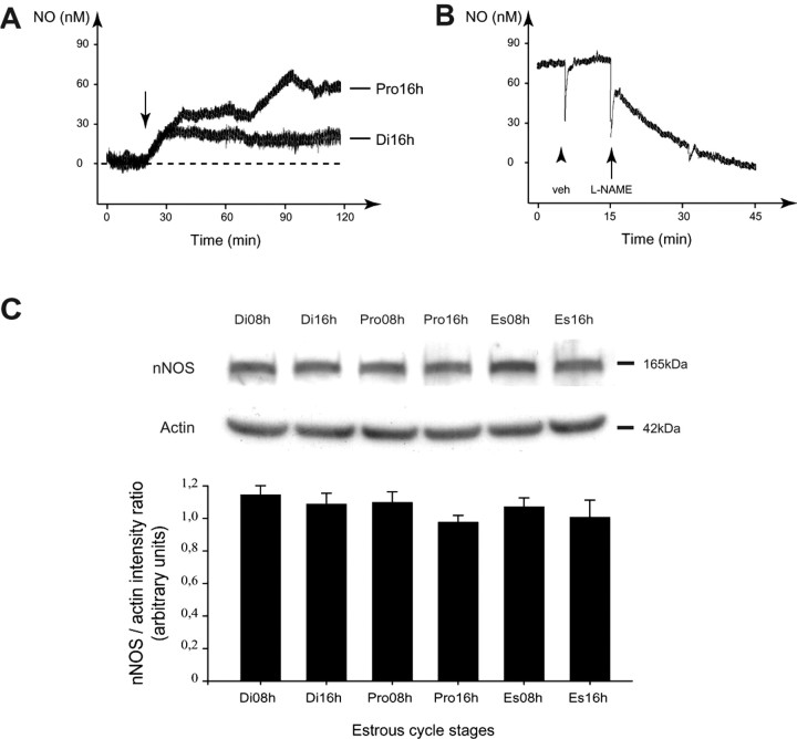
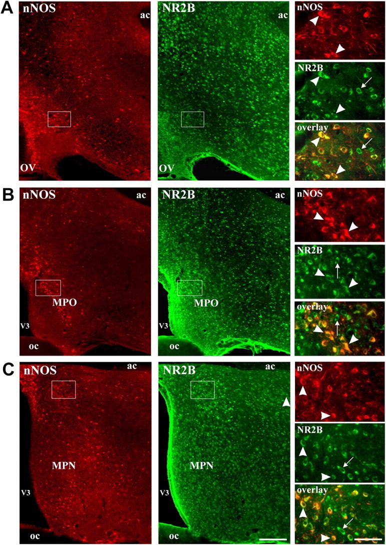
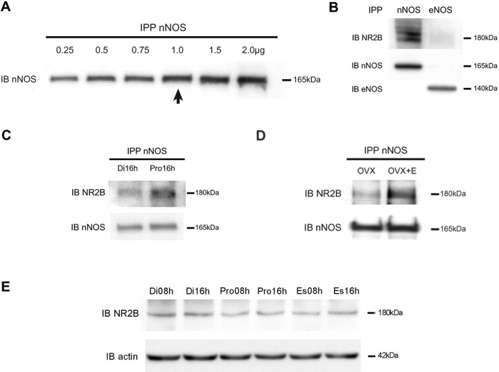
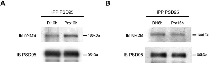
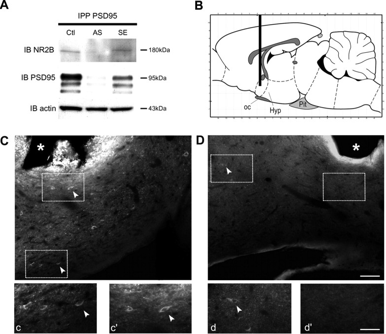
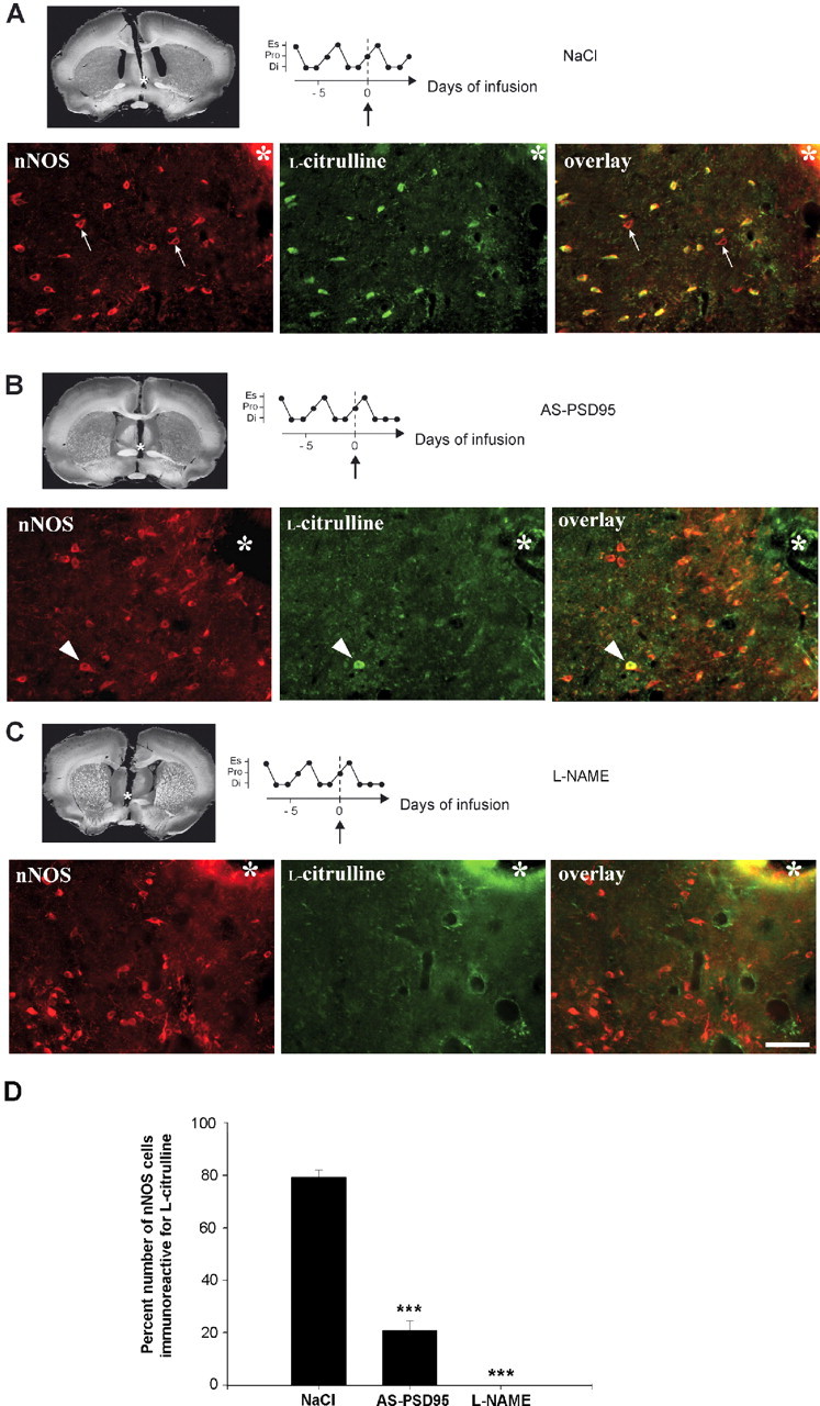
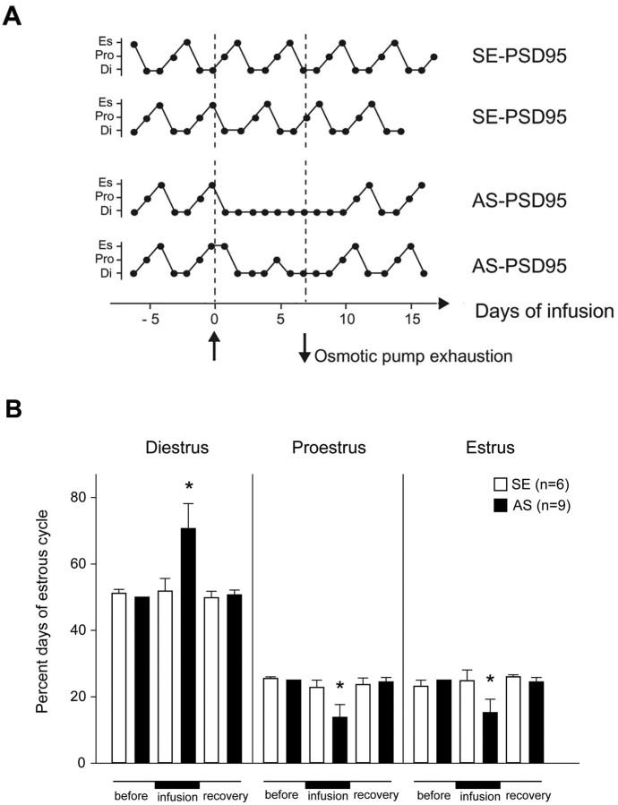
Considerable research has been devoted to the understanding of how nitric oxide (NO) influences brain function. Few studies, however, have addressed how its production is physiologically regulated. Here, we report that protein-protein interactions between neuronal NO synthase (nNOS) and glutamate NMDA receptors via the scaffolding protein postsynaptic density-95 (PSD-95) in the hypothalamic preoptic region of adult female rats is sensitive to cyclic estrogen fluctuation. Coimmunoprecipitation experiments were used to assess the physical association between nNOS and NMDA receptor NR2B subunit in the preoptic region of the hypothalamus. We found that nNOS strongly interacts with NR2B at the onset of the preovulatory surge at proestrus (when estrogen levels are highest) compared with basal-stage diestrous rats. Consistently, estrogen treatment of gonadectomized female rats also increases nNOS/NR2B complex formation. Moreover, endogenous fluctuations in estrogen levels during the estrous cycle coincide with changes in the physical association of nNOS to PSD-95 and the magnitude of NO release in the preoptic region. Finally, temporary and local in vivo suppression of PSD-95 synthesis by using antisense oligodeoxynucleotides leads to inhibition of nNOS activity in the preoptic region and disrupted estrous cyclicity, a process requiring coordinated activation of neurons containing gonadotropin-releasing hormone (the neuropeptide controlling reproductive function). In conclusion, our findings identify a novel steroid-mediated molecular mechanism that enables the adult mammalian brain to control NO release under physiological conditions.


Figures

Figure 1.
Figure 1.
NO effluxes generated by NOS across the estrous cycle. A, Representative profiles of spontaneous NO secretion from preoptic region explants at two different stages of the estrous cycle in the female rat. Differential current measured by the self-referencing probe, converted to flux (vertical axis; see Materials and Methods), increased after the addition of single preoptic explants in survival medium at the time indicated by the arrow. Comparisons between stages of the estrous cycle indicated that preoptic explants produced significantly more NO on the afternoon of proestrus than on the afternoon of diestrus (p = 0.014). B, Representative response of a proestrous rat preoptic explant to vehicle (veh; arrowhead) or to 1 mm L-NAME (arrow). Comparisons between treatments indicated that L-NAME, but not vehicle, significantly reduced NO production by the preoptic region during proestrus (p = 0.017). The transient drops in current on substance application (arrowhead and arrow) are artifacts. C, Absence of changes in nNOS protein expression in the preoptic region of the hypothalamus during the estrous cycle of the adult female rat. Proteins were collected from six different stages of the estrous cycle: diestrus II 8 h (Di08h) and 16 h (Di16h), proestrus 8 h (Pro08h) and 16 h (Pro16h), estrus 8 h (Es08h) and 16 h (Es16h). Twenty-five micrograms of protein per lane were electrophoresed and immunoblotted with antibodies to nNOS (top), and membranes were reprobed with antibodies to actin (middle). A representative blot from eight independent experiments is shown. Bottom, The protein levels are expressed in arbitrary densitometric units as the ratio between the nNOS signal and the signal obtained with constitutively expressed actin in each sample. Error bars indicate SEM. There is no statistically significant difference between groups.
Figure 2.
Figure 2.
NMDA receptor NR2B subunit is expressed by NO-producing neurons in the preoptic region. Representative illustration of nNOS-expressing neurons (red) and NR2B subunit detection (green) by fluorescent immunocytochemistry in coronal brain sections from female rats showing progressively caudal sections of the preoptic region containing the vascular organ of the lamina terminalis (OV) (A), the MPO (B), and the MPN (C). V3, Third ventricle; ac, anterior commissure; oc, optic chiasm. Right, High magnifications of the areas delineated by rectangles. Arrowheads indicate double-labeled neurons expressing nNOS and NMDA receptor NR2B subunit. Arrows indicate NR2B single-labeled neurons. Scale bars: low magnification, 210 μm; high magnification, 60 μm.
Figure 3.
Figure 3.
NMDA receptor NR2B subunit is highly physically associated with nNOS on the afternoon of proestrus. A, Protein (2200 μm) collected from the rat preoptic region was immunoprecipitated with increasing concentrations of specific nNOS antibodies, electrophoresed to size fractionate the immunoprecipitated species, and immunoblotted with the same antibody. To quantify coprecipitated species in the subsequent experiments, 1 μg of the nNOS antibody (arrow), which is a concentration that precipitates submaximal quantities of nNOS, was used to avoid any bias caused by potential variations in nNOS expression. B, One microgram of the rabbit polyclonal antibody to nNOS coprecipitated NR2B from female rat preoptic region protein extracts, whereas 1 μg of the rabbit polyclonal antibody to eNOS failed to do so. C, Increased association of nNOS with NR2B on the afternoon of proestrus (Pro16h) in the preoptic region of adult cycling female rats. D, Estradiol (E) promotes nNOS/NR2B complex formation in the preoptic region of the female rat. A representative blot illustrating the increase in nNOS/NR2B coimmunoprecipitation in the OVX+E female rat is shown. E, Representative blot showing absence of changes in NR2B protein expression in the preoptic region of the hypothalamus during the estrous cycle of the adult female rat. The effective amount of protein loaded is represented by actin blot. Proteins were collected and processed as detailed in Figure 1C. IPP, Immunoprecipitated; IB, immunoblotted.
Figure 4.
Figure 4.
Differential NR2B/nNOS complex formation involves changes in physical association of PSD-95/nNOS but not NR2B/PSD-95. Immunoprecipitation (IPP) of preoptic region proteins with antibodies against PSD-95 results in the coprecipitation of nNOS (A) and of NR2B (B). Each observation derives from preoptic region tissue pooled from four rats. IB, Immunoblot.
Figure 5.
Figure 5.
Use of PSD-95 antisense ODNs to disrupt PSD-95 expression in neurons of the rat preoptic region. A, Selective decrease in PSD-95 expression and its association with NR2B in hypothalamic neurons treated with the antisense ODN to PSD-95 in vitro. Cells were treated with the antisense ODN (5 μm) or a sense sequence for 7 d, and protein extracts were immunoprecipitated (IPP) with antibodies to PSD-95 and sequentially immunoblotted (IB) with NR2B and PSD-95 antibodies. The supernatant resulting from immunoprecipitation was subjected to straight Western blotting for actin. Ctl, Control; AS, antisense; SE, sense. B–D, Central administration of an antisense PSD-95 ODN extinguishes PSD-95 immunoreactivity at the infusion site. B, Schematic diagram representing the stereotaxically implanted 28 gauge infusion cannula into the preoptic region of cycling female rats. ODNs (0.2 nmol/μl) were delivered via a stereotaxically implanted stainless steel 28 gauge cannula connected to a subcutaneously implanted osmotic pump delivering its contents at a rate of 0.5 μl/h for up to 7 d. oc, Optic chiasm; Hyp, hypothalamus; Pit, pituitary. C, D, Representative illustration of PSD-95 detection by fluorescent immunocytochemistry in coronal brain sections at the tip of the implantation site in sense PSD-95 ODN-infused animals (C) or in antisense PSD-95-treated rats (D). c, c′, d, d′, Higher magnifications of the areas delineated by rectangles. The asterisk indicates the implanted cannula tip; arrowheads indicate PSD-95-immunoreactive neuronal cell bodies. Scale bars: low magnification, 70 μm; high magnification, 35 μm.
Figure 6.
Figure 6.
Central administration of an antisense PSD-95 (AS-PSD-95) ODN impairs NO formation in hypothalamic nNOS neurons. A–C, Antisense PSD-95 ODN (B), L-NAME (5 mm, a NOS inhibitor) (C), or vehicle (0.9% NaCl) (A) was delivered into the preoptic region (top left) of adult female rats with regular estrous cycles as described in Figure 7; nNOS catalytic activity was evaluated in vivo by staining for l-citrulline (green), which is formed by nNOS (red) stoichiometrically with NO. In parallel, the effect of infusion on estrous cyclicity was monitored (top right). Infusion starts at day 0 (upward arrow), and animals were killed on day 4, before the pump content was exhausted. A representative animal of each treatment group is shown. The asterisks indicate cannula implantation sites, arrows indicate nNOS single-labeled neurons, and arrowheads indicate a nNOS-expressing neuron immunolabeled for l-citrulline. Di, Diestrus; Pro, proestrus; Es, estrus. Scale bar, 70 μm. D, Bar graph illustrating the percentage number of nNOS neurons immunoreactive for l-citrulline at the tip of the infusion site from animals treated with NaCl, AS-PSD-95 ODNs, or L-NAME (***p < 0.001 compared with all other groups, one-way ANOVA; n = 4–5 each).
Figure 7.
Figure 7.
Central administration of an antisense PSD-95 ODN results in estrous cycle disruption in sexually mature rats. A, Representative estrous cycle profiles showing disruption of estrous cyclicity in young adult rats by the infusion of antisense PSD-95 (AS-PSD-95) but not of sense PSD-95 (SE-PSD-95) ODNs (0.2 nmol/μl each) into the hypothalamic preoptic region. Infusion started at day 0 (upward arrow) and ended 7 d later (downward arrow), when the pump content was exhausted. Animals were allowed to survive for 1 additional week (recovery) after pump exhaustion. Di, Diestrus; Pro, proestrus; Es, estrus. B, Quantitative analysis of the alterations in estrous cyclicity caused by antisense PSD-95-ODN infusion into the hypothalamic preoptic region. *p < 0.05 compared with before and recovery for the same stage and the same treatment (repeated-measures ANOVA). Error bars indicate SEM. The number of independent observations per group is shown in parentheses. AS, antisense; SE, sense.

Similar articles

Cited by

References

    1. Aarts M, Liu Y, Liu L, Besshoh S, Arundine M, Gurd JW, Wang YT, Salter MW, Tymianski M. Treatment of ischemic brain damage by perturbing NMDA receptor- PSD-95 protein interactions. Science. 2002;298:846–850. - PubMed
    1. Akama KT, McEwen BS. Estrogen stimulates postsynaptic density-95 rapid protein synthesis via the Akt/protein kinase B pathway. J Neurosci. 2003;23:2333–2339. - PMC - PubMed
    1. Altschul SF, Madden TL, Schaffer AA, Zhang J, Zhang Z, Miller W, Lipman DJ. Gapped BLAST and PSI-BLAST: a new generation of protein database search programs. Nucleic Acids Res. 1997;25:3389–3402. - PMC - PubMed
    1. Amateau SK, McCarthy MM. A novel mechanism of dendritic spine plasticity involving estradiol induction of prostaglandin-E2. J Neurosci. 2002;22:8586–8596. - PMC - PubMed
    1. Beauvillain JC, Tramu G. Immunocytochemical demonstration of LH-RH, somatostatin, and ACTH-like peptide in osmium-postfixed, resin-embedded median eminence. J Histochem Cytochem. 1980;28:1014–1017. - PubMed

Publication types

Substances

LinkOut - more resources