Comparative Study
. 2007 Jun 6;27(23):6115-27.
doi: 10.1523/JNEUROSCI.0180-07.2007.

Nuclear factor I coordinates multiple phases of cerebellar granule cell development via regulation of cell adhesion molecules

Affiliations
Comparative Study

Nuclear factor I coordinates multiple phases of cerebellar granule cell development via regulation of cell adhesion molecules

Wei Wang et al. J Neurosci. .

Abstract

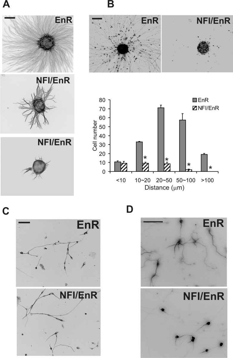
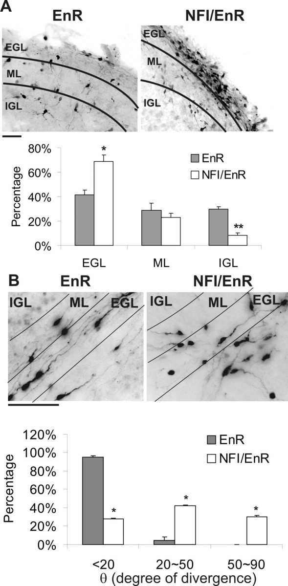
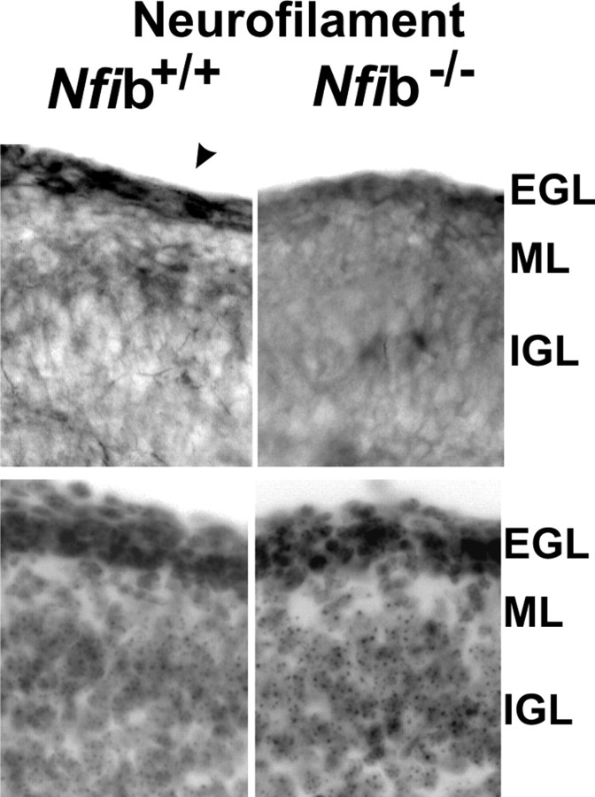
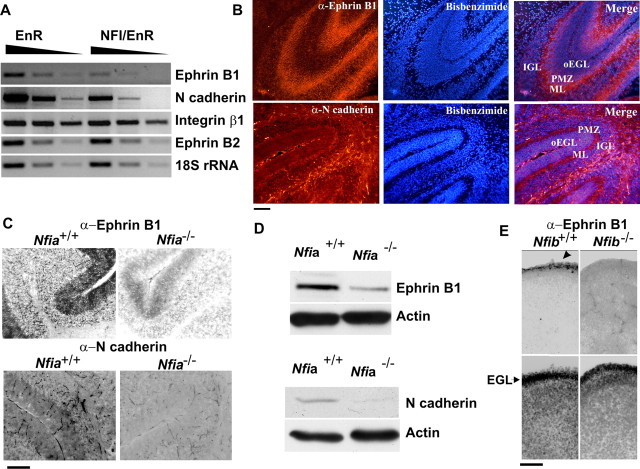
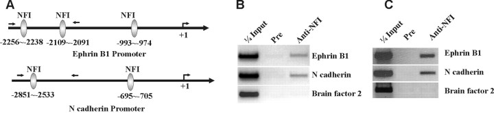
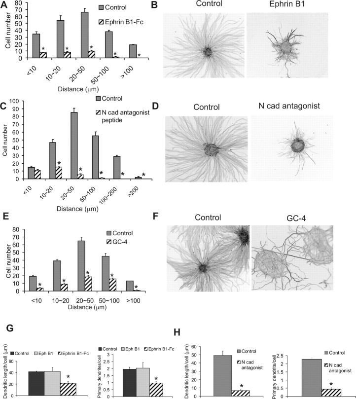
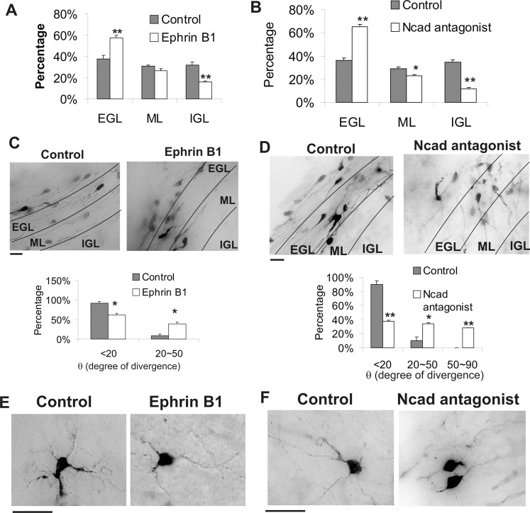
A central question is how various stages of neuronal development are integrated as a differentiation program. Here we show that the nuclear factor I (NFI) family of transcriptional regulators is expressed and functions throughout the postmitotic development of cerebellar granule neurons (CGNs). Expression of an NFI dominant repressor in CGN cultures blocked axon outgrowth and dendrite formation and decreased CGN migration. Inhibition of NFI transactivation also disrupted extension and fasciculation of parallel fibers as well as CGN migration to the internal granule cell layer in cerebellar slices. In postnatal day 17 Nfia-deficient mice, parallel fibers were greatly diminished and disoriented, CGN dendrite formation was dramatically impaired, and migration from the external germinal layer (EGL) was retarded. Axonal marker expression also was disrupted within the EGL of embryonic day 18 Nfib-null mice. NFI regulation of axon extension was observed under conditions of homotypic cell contact, implicating cell surface proteins as downstream mediators of its actions in CGNs. Consistent with this, the cell adhesion molecules ephrin B1 and N-cadherin were identified as NFI gene targets in CGNs using inhibitor and Nfi mutant analysis as well as chromatin immunoprecipitation. Functional inhibition of ephrin B1 or N-cadherin interfered with CGN axon extension and guidance, migration, and dendritogenesis in cell culture as well as in situ. These studies define NFI as a key regulator of postmitotic CGN development, in particular of axon formation, dendritogenesis, and migratory behavior. Furthermore, they reveal how a single transcription factor family can control and integrate multiple aspects of neuronal differentiation through the regulation of cell adhesion molecules.


Figures

Figure 1.
Figure 1.
Expression of NFI family members in the P6 mouse cerebellum. Immunofluorescence for NFIA, NFIB, and NFIX are shown. In each case, specific nuclear staining in CGNs first appears within the deeper EGL (PMZ) and persists as cells migrate through the ML and then populate the IGL. NFIC antibody did not exhibit specific staining (data not shown). Scale bar, 50 μm.
Figure 2.
Figure 2.
NFI regulates process formation and migration. A, The NFI dominant repressor impairs neurite outgrowth from CGN reaggregates. CGNs were infected with lentivirus expressing HA-tagged NFI/EnR or EnR during cell reaggregation and examined 20 h after plating for neurite extension using β-III tubulin immunostaining. B, NFI is required for migration of CGNs from reaggregates. Top, Reaggregates were stained for HA to identify transduced CGN nuclei. Bottom, The distribution of HA+ neurons migrating from reaggregates. C, The NFI dominant repressor does not significantly affect neurite outgrowth in fully dissociated CGN cultures. Dissociated CGNs were infected with NFI/EnR or EnR virus on 0 DIV and stained for β-III tubulin on 2 DIV. D, NFI is required for dendrite formation in dissociated CGN cultures. CGNs were transduced with NFI/EnR or EnR lentiviruses and then stained for MAP2. Both dendrite length and number per cell were significantly reduced by the NFI dominant repressor relative to control. *p < 0.001, significantly different from control. Scale bars: A–C, 100 μm; D, 50 μm.
Figure 3.
Figure 3.
NFI transactivation is required for parallel fiber extension and CGN migration in situ. A, The dominant repressor impairs CGN migration from the EGL to the IGL. Top, Representative images of GFP+ CGNs infected with NFI/EnR or EnR retroviruses in sagittal cerebellar slices. Bottom, The percentage of GFP+ cells present within the EGL, ML, and IGL expressed as the mean. *p < 0.05, **p < 0.001, significantly different from EnR control. B, Parallel fiber extension and orientation are markedly altered by inhibition of NFI. Top, GFP+ immunostaining of parallel fibers within the EGL/ML region of coronal cerebellar slices transduced with NFI/EnR or EnR retroviruses. Essentially all GFP+ cells detected within this region contained bipolar processes typical of forming parallel fibers. Note that labeled cells lying below the indicated EGL region derive from an apposing EGL. Bottom, The angle of divergence (θ) from the parallel orientation, expressed in degrees, for GFP+ axons. *p < 0.001, significantly different from control. Scale bars, 50 μm.
Figure 4.
Figure 4.
Abnormal cerebellar development in Nfi-null mice. A, Bisbenzimide staining of sagittal sections through the vermis of P17 mice showing altered foliation in Nfi mutants. Top, Nfia+/+. Bottom, Nfia−/−. Lobules are labeled with roman numerals. B, DiI-labeled parallel fibers within cerebellar coronal sections from wild-type and Nfia-null animals. Left, Arrowheads indicate parallel fibers within the ML extending away from the DiI crystal inserted at the midline. Axon extension is reduced throughout the cerebellar vermis of Nfia-null mice compared with wild-type mice. Right, Higher magnification of the ML showing dramatically shortened axons in Nfia-null mice. Note the parallel orientation of fibers in wild-type cerebella. Bottom, Numerous disorientated axons (arrowheads) extending toward the IGL and/or meninges are observed only in Nfia-null cerebella. C, DiI-labeled dendritic processes on CGNs within the IGL of the anterior vermis of wild-type (top) and Nfia-null (bottom) mice. Wild-type CGNs exhibited typical claw-shaped dendrites (arrowheads), which were lacking on CGNs within the anterior cerebella of mutant mice. Note that the intensity of DiI staining of CGN cell bodies within the IGL was identical in wild-type and Nfia mutant mice. D, P17 Nfia-null mice exhibit a residual EGL containing differentiating CGNs. Left, NeuN+ cells within sagittal sections of wild-type and Nfia−/− mice from lobule VI (regions are indicated by rectangles in A). Arrowheads indicate ectopic cells within the ML of mutant mice. Right, Bisbenzimide staining of the same sections. E, Left, Calbindin immunostaining of Purkinje cells in wild-type and Nfia-null cerebella. The location and densities of these cells were the same in both genotypes. Dendritic processes in Nfia mutant mice were less extensive along their superficial aspect (arrowhead) possibly attributable to reduced parallel fiber extension and residual EGL cells within this region. Right, Bisbenzimide staining of the same sections showing the residual EGL in Nfia mutant cerebellum. Scale bars: B–E, 50 μm. All images derive from the vermis of P17 cerebella.
Figure 5.
Figure 5.
Axon formation is altered in the late embryonic cerebellum of Nfib-null mice. Top, Sagittal sections of E18 cerebella from Nfib+/+ and Nfib−/− mice stained for phospho-neurofilament, which is greatly reduced in Nfib mutant mice. The posterior cerebellum is shown. Arrowhead indicates immunoreactive cells within the EGL of wild-type cerebellum. Bottom, Bisbenzimide staining of the same sections. Scale bar, 50 μm.
Figure 6.
Figure 6.
Expression of cell adhesion molecules is regulated by NFI in CGNs. A, Ephrin B1 and N-cadherin transcripts are specifically reduced by the NFI dominant repressor in CGNs. Semiquantitative RT-PCR analysis of cell adhesion or guidance molecule mRNAs in CGN cultures transduced with NFI/EnR or EnR lentiviruses. Samples were analyzed using threefold serial dilutions. 18S ribosomal RNA was assayed as a loading control. B, Immunostaining for ephrin B1 (top) and N-cadherin (bottom) in the vermis of P6 mouse cerebella. oEGL, Outer EGL region. C, Cell adhesion molecule immunostaining is reduced in Nfia mutant mice. Sagittal sections through the vermis of P17 Nfia+/+ and Nfia−/− mice were stained with antibodies against ephrin B1 (top) and N-cadherin (bottom). D, Western blot analysis of ephrin B1 and N-cadherin in mouse cerebella showing reduced expression of these proteins in Nfia-deficient mice. E, Ephrin B1 immunostaining (top) is reduced in E18 Nfib−/− mouse cerebellum relative to Nfib+/+ controls. The arrowhead indicates labeled cells within the EGL of wild-type mice. Bottom, Bisbenzimide staining of the same sections. Scale bars, 50 μm.
Figure 7.
Figure 7.
In vivo binding of NFI to cell adhesion molecule promoters in postmitotic CGNs. A, Locations of consensus NFI binding sites within the mouse ephrin B1 and N-cadherin promoters. The approximate positions of PCR primers within the 5′ regions for ChIP assays are indicated by arrows. B, ChIP assay showing that NFI proteins are bound in vivo to the ephrin B1 and N-cadherin promoter regions in 6 DIV CGN cell cultures. C, NFI binds to the ephrin B1 and N-cadherin promoters in intact mouse cerebellar tissue. ChIP assay using nuclei prepared from P15 cerebella, >85% of which derive from CGNs based on NeuN and NeuroD immunostaining. Mouse brain factor 2 genomic sequences were used as a negative control. Pre, Preimmune serum.
Figure 8.
Figure 8.
Inhibitors of ephrin B and N-cadherin impair CGN neurite outgrowth, migration, and dendrite formation. CGN cultures were treated for 24 h with 50 μg/ml ephrin B1-Fc or BSA (A, B, G), 400 μg/ml N-cadherin (Ncad) peptide antagonists or control peptide (C, D, H), and 50 μg/ml GC-4 or IgG isotype control antibodies (E, F). A, C, E, Antagonists inhibit CGN migration from reaggregates. Bisbenzimide staining of cell nuclei was used to identify migrated cells. B, D, F, Neurites extension is inhibited by N-cadherin and ephrin B antagonists. Neurites were identified using anti-β III tubulin staining. G, H, Inhibition of dendritic length (left) and the number of primary dendrites per cell (right) by ephrin B1-Fc and N-cadherin antagonist peptides using MAP2 antibody. Soluble EphB1-Fc also was tested at 50 μg/ml in G. *p < 0.001, significantly different from control values in all cases except in G (p < 0.01).
Figure 9.
Figure 9.
Cell adhesion molecule inhibitors disrupt differentiation of CGNs in situ. A, B, CGN radial migration is inhibited by cell adhesion molecule antagonists. Cerebellar slices were transduced with GFP retroviruses and treated with 50 μg/ml ephrinB1-Fc, 400 μg/ml N-cadherin inhibitor peptides, or control reagents for 60 h. Distributions of viral-transduced GFP+ cells within different cerebellar layers are shown. Both inhibitors significantly reduced CGN migration relative to controls. *p < 0.05; **p < 0.01. C, D, Top, Antagonists inhibit parallel fiber extension and induce axon misorientation. GFP+ parallel fibers in the PMZ/ML region of coronal slices treated with 50 μg/ml ephrinB1-Fc or 400 μg/ml N-cadherin inhibitor peptides for 48 h. Labeled cells lying outside the indicated layers derive from an adjacent EGL (see legend to Fig. 3B). Bottom, The orientation of GFP+ axons was significantly altered by the soluble inhibitors relative to controls. *p < 0.01; **p < 0.001. θ, Angle of divergence from the parallel orientation for GFP+ axons, expressed in degrees. E, F, Antagonists inhibit extension of GFP-labeled dendrites from retroviral-transduced CGNs within the IGL of cerebellar slices. Cultures were treated with either ephrinB1-Fc (E) or N-cadherin antagonist peptides (F) or control reagents for 60 h.

Similar articles

Cited by

References

    1. Altman J. Postnatal development of the cerebellar cortex in the rat. 3. Maturation of the components of the granular layer. J Comp Neurol. 1972;145:465–513. - PubMed
    1. Arregui C, Pathre P, Lilien J, Balsamo J. The nonreceptor tyrosine kinase fer mediates cross-talk between N-cadherin and beta1-integrins. J Cell Biol. 2000;149:1263–1274. - PMC - PubMed
    1. Aruga J, Minowa O, Yaginuma H, Kuno J, Nagai T, Noda T, Mikoshiba K. Mouse Zic1 is involved in cerebellar development. J Neurosci. 1998;18:284–293. - PMC - PubMed
    1. Aruga J, Inoue T, Hoshino J, Mikoshiba K. Zic2 controls cerebellar development in cooperation with Zic1. J Neurosci. 2002;22:218–225. - PMC - PubMed
    1. Barami K, Kirschenbaum B, Lemmon V, Goldman SA. N-cadherin and Ng-CAM/8D9 are involved serially in the migration of newly generated neurons into the adult songbird brain. Neuron. 1994;13:567–582. - PubMed

Publication types

Substances

LinkOut - more resources