

Where am I now? Distinct roles for parahippocampal and retrosplenial cortices in place recognition
- PMID: 17553986
- PMCID: PMC6672165
- DOI: 10.1523/JNEUROSCI.0799-07.2007
Where am I now? Distinct roles for parahippocampal and retrosplenial cortices in place recognition
Abstract
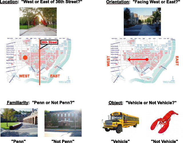
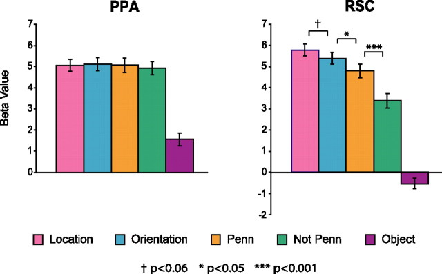
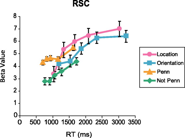
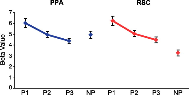
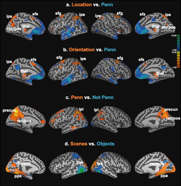
A key component of spatial navigation is the ability to use visual information to ascertain where one is located and how one is oriented in the world. We used functional magnetic resonance imaging to examine the neural correlates of this phenomenon in humans. Subjects were scanned while retrieving different kinds of topographical and nontopographical information in response to visual scenes. In the three critical conditions, they viewed images of a familiar college campus, and reported either the location of the place depicted in the image (location task), the compass direction that the camera was facing when the image was taken (orientation task), or whether the location was on campus or not (familiarity task). Our analyses focused on the retrosplenial cortex (RSC)/parietal-occipital sulcus region and the parahippocampal place area (PPA), which previous studies indicate play a critical role in place recognition. RSC activity depended on the type of information retrieved, with the strongest response in the location task. In contrast, PPA activity did not depend on the retrieval task. Additional analyses revealed a strong effect of familiarity in RSC but not in the PPA, with the former region responding much more strongly to images of the familiar campus than to images of an unfamiliar campus. These results suggest that the PPA and RSC play distinct but complementary roles in place recognition. In particular, the PPA may primarily support perception of the immediate scene, whereas RSC may support memory retrieval mechanisms that allow the scene to be localized within the broader spatial environment.
Figures
Similar articles
-
Parahippocampal and retrosplenial contributions to human spatial navigation.Trends Cogn Sci. 2008 Oct;12(10):388-96. doi: 10.1016/j.tics.2008.07.004. Epub 2008 Aug 28. Trends Cogn Sci. 2008. PMID: 18760955 Free PMC article. Review.
-
A Posterior-Anterior Distinction between Scene Perception and Scene Construction in Human Medial Parietal Cortex.J Neurosci. 2019 Jan 23;39(4):705-717. doi: 10.1523/JNEUROSCI.1219-18.2018. Epub 2018 Nov 30. J Neurosci. 2019. PMID: 30504281 Free PMC article.
-
Dissociable Neural Systems for Recognizing Places and Navigating through Them.J Neurosci. 2018 Nov 28;38(48):10295-10304. doi: 10.1523/JNEUROSCI.1200-18.2018. Epub 2018 Oct 22. J Neurosci. 2018. PMID: 30348675 Free PMC article.
-
Spatial frequency processing in scene-selective cortical regions.Neuroimage. 2015 May 15;112:86-95. doi: 10.1016/j.neuroimage.2015.02.058. Epub 2015 Mar 6. Neuroimage. 2015. PMID: 25754068
-
Human spatial navigation: Neural representations of spatial scales and reference frames obtained from an ALE meta-analysis.Neuroimage. 2021 Sep;238:118264. doi: 10.1016/j.neuroimage.2021.118264. Epub 2021 Jun 12. Neuroimage. 2021. PMID: 34129948 Review.
Cited by
-
Retrosplenial Cortical Neurons Encode Navigational Cues, Trajectories and Reward Locations During Goal Directed Navigation.Cereb Cortex. 2017 Jul 1;27(7):3713-3723. doi: 10.1093/cercor/bhw192. Cereb Cortex. 2017. PMID: 27473323 Free PMC article.
-
Route and survey processing of topographical memory during navigation.Psychol Res. 2010 Nov;74(6):545-59. doi: 10.1007/s00426-010-0276-5. Epub 2010 Feb 20. Psychol Res. 2010. PMID: 20174930
-
Recruitment of Control and Representational Components of the Semantic System during Successful and Unsuccessful Access to Complex Factual Knowledge.J Neurosci. 2022 Jun 15;42(24):4879-4890. doi: 10.1523/JNEUROSCI.2485-21.2022. Epub 2022 May 12. J Neurosci. 2022. PMID: 35552235 Free PMC article.
-
Retrosplenial cortex in spatial memory: focus on immediate early genes mapping.Mol Brain. 2021 Dec 4;14(1):172. doi: 10.1186/s13041-021-00880-w. Mol Brain. 2021. PMID: 34863215 Free PMC article. Review.
-
Evidence for the Concreteness of Abstract Language: A Meta-Analysis of Neuroimaging Studies.Brain Sci. 2021 Dec 28;12(1):32. doi: 10.3390/brainsci12010032. Brain Sci. 2021. PMID: 35053776 Free PMC article. Review.
References
-
- Aguirre GK, D'Esposito M. Topographical disorientation: a synthesis and taxonomy. Brain. 1999;122:1613–1628. - PubMed
-
- Aguirre GK, Detre JA, Alsop DC, D'Esposito M. The parahippocampus subserves topographical learning in man. Cereb Cortex. 1996;6:823–829. - PubMed
-
- Aminoff E, Gronau N, Bar M. The parahippocampal cortex mediates spatial and nonspatial associations. Cereb Cortex. 2007 in press. - PubMed
-
- Bar M, Aminoff E. Cortical analysis of visual context. Neuron. 2003;38:347–358. - PubMed
-
- Burgess N. Spatial memory: how egocentric and allocentric combine. Trends Cogn Sci. 2006;10:551–557. - PubMed
Publication types
MeSH terms
Grants and funding
LinkOut - more resources
Full Text Sources