Comparative Study
. 2007 Jun 6;27(23):6150-62.
doi: 10.1523/JNEUROSCI.1466-07.2007.

UNC-31 (CAPS) is required for dense-core vesicle but not synaptic vesicle exocytosis in Caenorhabditis elegans

Affiliations
Comparative Study

UNC-31 (CAPS) is required for dense-core vesicle but not synaptic vesicle exocytosis in Caenorhabditis elegans

Sean Speese et al. J Neurosci. .

Abstract

Previous studies indicated that CAPS (calcium-dependent activator protein for secretion) functions as an essential component for the Ca2+-dependent exocytosis of dense-core vesicles in neuroendocrine cells. However, recent mouse knock-out studies suggested an alternative role in the vesicular uptake or storage of catecholamines. To genetically assess the functional role of CAPS, we characterized the sole Caenorhabditis elegans CAPS ortholog UNC-31 (uncoordinated family member) and determined its role in dense-core vesicle-mediated peptide secretion and in synaptic vesicle recycling. Novel assays for dense-core vesicle exocytosis were developed by expressing a prepro-atrial natriuretic factor-green fluorescent protein fusion protein in C. elegans. unc-31 mutants exhibited reduced peptide release in vivo and lacked evoked peptide release in cultured neurons. In contrast, cultured neurons from unc-31 mutants exhibited normal stimulated synaptic vesicle recycling measured by FM4-64 [N-(3-triethylammoniumpropyl)-4-(6-(4-diethylamino)phenyl)hexatrienyl)pyridinium dibromide] dye uptake. Conversely, UNC-13, which exhibits sequence homology to CAPS/UNC-31, was found to be essential for synaptic vesicle but not dense-core vesicle exocytosis. These findings indicate that CAPS/UNC-31 function is not restricted to catecholaminergic vesicles but is generally required for and specific to dense-core vesicle exocytosis. Our results suggest that CAPS/UNC-31 and UNC-13 serve parallel and dedicated roles in dense-core vesicle and synaptic vesicle exocytosis, respectively, in the C. elegans nervous system.


Figures

Figure 1.
Figure 1.
Genomic structure, mutant allele characterization, and protein architecture for UNC-31. A, Schematic representation of the UNC-31 genomic structure. For details for unc-31 mutant alleles, see Table 1. All alleles are nonsense or splice site mutations except ox299, e714, and e928. ox299 corresponds to the missense mutation I530K and e714 corresponds to the missense mutation G538E. e928 is a deletion indicated by solid black line; the double bar underneath represents a transposition of a fragment of the unc-31 locus detected in the e928 allele. B, UNC-31 protein architecture. Identified domains in the protein depicted are as follows: coiled-coil (residues 133–159); 20 aa domain defined by alleles u377 and sa534 (273–293) (Table 1); C2 domain (460–558); and PH domain (582–689C). The coiled-coil, C2, and PH domain were defined by Simple Modular Architecture Research Tool. The MUN domain (730–1180) (Basu et al., 2005) was defined by multiple alignment programs. The dense-core vesicle binding domain (DCVBD) (1181 end) was defined based on alignment and Grishanin et al. (2002). C, Western blotting for UNC-31. Homogenates (100 μg of protein) from wild-type (WT) and mutant strains were analyzed by Western blotting with an affinity-purified UNC-31 antibody. Results shown are representative of at least two independent experiments. unc-31(e714) exhibited decreased UNC-31 levels (22 ± 13% of wild type, mean ± SE; n = 3). D, Alignment of protein sequences for a portion of the C2 domain of C. elegans UNC-31 with those from human, mouse, and Drosophila orthologs. Highlighted in the red box are the residues mutated in unc-31(ox299) and unc-31(e714). E, Schematic representation of unc-31::GFP transcriptional and translational constructs and an overlapping fragment that was coinjected. Coinjection was done to provide additional promoter elements that were necessary for full rescue (S. Speese, unpublished observations). UTR, Untranslated region; NLS, nuclear localization sequence.
Figure 2.
Figure 2.
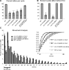
Defecation and movement in unc-31. A, Graph showing the percentage of cycles in which an aBoc was observed in a particular genotype. The number of aBoc events was divided by the total number of cycles scored to generate the percentages. B, The number of cycles with an aBoc before an Emc was divided by the number of cycles that contained both an aBoc and an Emc. All worms were scored blind for 10 cycles each. C, Histogram showing the analysis of unc-31(e928) movement on and off food. Velocity (millimeters per second) of the worms was measured for 45 min using a video tracking system and computer analysis. Speeds between 0 and 0.02 mm/s were equivalent to the worm not moving. Inset is the same data graphed as a cumulative plot. All animals in these quantitative phenotypic studies were assayed as young adults. In the movement assays, prestarved unc-31(e928) animals were picked as L4s and transferred to a plate without a bacterial lawn for 5 h. Immediately after the starvation period, they were tracked on an assay plate without food. WT, Wild type.
Figure 3.
Figure 3.
UNC-31 is expressed in neurons and is enriched at synapses. A, Confocal image of an adult hermaphrodite expressing GFP under the unc-31 promoter (oxEx608[Punc-31:GFP]). Expression of unc-31 is pan-neuronal with additional expression in the spermatheca and vulval muscles. Anterior is left. B, Confocal image of the ventral surface of an adult hermaphrodite expressing GFP under the unc-31 promoter. Expression is observed in the vulval muscles VM1 and VM2. Expression is also seen in the UV1 cells. C, Confocal images of unc-31(e928) oxEx608[UNC-31:GFP]. UNC-31:GFP protein localizes to the synaptic-rich region of the nerve ring as well as the dorsal and ventral nerve cords. GFP expression inside the dashed circle around the posterior bulb of the pharynx can be disregarded because it arises from the coinjection marker (Pmyo-2:HIS::GFP). Additional diffuse expression of the coinjection marker can also be seen in the isthmus of the pharynx. D, Subcellular localization of UNC-31:GFP in the SAB neurons of the head. Punctate expression as well as colocalization with the synaptic vesicle marker synaptobrevin (α-SNB-1) indicate that UNC-31 is localized to synaptic terminals, reported previously by Charlie et al. (2006).
Figure 4.
Figure 4.
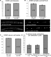
Absence of developmental defects in unc-31. A, Quantification of GABA motor neuron outgrowth indicates there are no defects in pathfinding in unc-31(u280). Confocal Z-series were captured of each animal at 40×, and the numbers of axons that crossed the midline and terminated on the dorsal nerve cord were counted. B, Synaptogenesis was evaluated by using GFP-tagged synaptobrevin (SNB-1:GFP). Quantification of SNB-1:GFP puncta density indicates that unc-31 mutants have normal numbers of GABA motor neuron synapses in the dorsal nerve cord. Puncta density was determined for a single animal by taking images of the dorsal nerve cord with a 63× lens at the posterior and anterior reflexes of the gonad. Puncta density in these two images was averaged to get the puncta per 10 μm. C, By counting the number of GFP-positive cells in a strain expressing GFP in GABA neurons (EG1846), it was determined that all GABA neurons are properly specified in unc-31 mutants. D, Acute expression of unc-31 from a heat shock (HS) promoter is able to rescue the anterior body contraction defect in unc-31(e928). Six cycles were observed for each animal scored to determine the percentage of cycles that had a contraction. All animals in these developmental assays were analyzed as young adults.
Figure 5.
Figure 5.
UNC-31 but not UNC-13 is essential for evoked dense-core vesicle exocytosis in cultured neurons. A, Images of the tgIs5[Paex-3:ANF::GFP] strain in either wild-type (WT), unc-31(e928), or unc-104(e1265) backgrounds. Images are of adult hermaphrodites, and anterior is up. nr, Nerve ring; vnc, ventral nerve cord; dnc, dorsal nerve cord. Scale bar, 25 μm. B, Sucrose velocity sedimentation of C. elegans dense-core vesicles and synaptic vesicles. Dense-core vesicles and synaptic vesicles were prepared from tgIs5[Paex-3:ANF::GFP] and jsIs219[synaptogyrin::GFP] strains, respectively, and analyzed on sucrose gradients for corresponding ANF–GFP (♦) or synaptogyrin–GFP (■) in the top. Western blotting was used to detect mature IDA-1 (■) or IDA-1 precursor (♦) in the bottom. C, Image of a cultured neuron from the tgIs5[Paex-3:ANF::GFP] strain in which the punctuate localization of ANF–GFP in cell bodies and neurites is shown. Scale bar, 2 μm. D, Time lapse images of ANF–GFP content in nerve cell bodies. Cultured neurons derived from tgIs5 (wild type), tgIs5 unc-31(e928), and tgIs5 unc-13(s69) strains were imaged immediately after addition of stimulation buffer and at 30 min and 60 min. E, Quantitation of ANF–GFP fluorescence in cultured neurons. Fluorescence in cell bodies was quantitated for wild-type neurons incubated in resting buffer (n = 14) (×) and in depolarization buffer for wild-type (n = 25) (♦), unc-31(e928) (n = 29) (■), or unc-13(s69) (n = 24) (▴) neurons. Mean ± SEM values of percentage release correspond to percentage fluorescence loss from cell bodies.
Figure 6.
Figure 6.
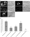
unc-31 mutants release less ANF–GFP into the body cavity. A, For each strain, the left represents a maximum pixel intensity projection of the GFP signal arising from ANF–GFP that has been taken up into endocytic compartments of the coelomocytes. The yellow dashed line indicates the location of the coelomocytes, which are shown in DIC images on the right. Fluorescence outside of the yellow dashed line is gut autofluorescence. B, Graph showing the normalized total pixel intensity per coelomocyte (CC). The total pixel intensity per coelomocyte of ANF–GFP in wild-type EG3680 worms was set to an arbitrary fluorescence unit (A.U.) of 100 to enable comparison of separate experiments in which the wild-type strain EG3680 oxIs206[ANF:GFP] was compared with mutant strains EG3681 unc-31(e928) oxIs206[ANF:GFP], EG3682 unc-31(u280) oxIs206[ANF:GFP]), EG3345 unc-13(s69) oxIs206[ANF:GFP], and EG3683 unc-13(e51) oxIs206[ANF:GFP]. InSpeck calibration microspheres (Invitrogen) were used to calibrate output of the microscope for fluorescence normalization. The SEM for the control strain (EG3680) was calculated as a weighted (based on total n) average of the SEM from three separate experiments, which differed by <8%. Statistical analyses were conducted for data within an experiment, and all genotypes were singly compared with control via an unpaired t test (**p = 0.002, ***p < 0.0001). The complete lack of ANF:GFP in coelomocytes of unc-13(s69) mutants is only reflective of the analyzed posterior coelomocytes, whereas anterior coelomocytes contained some ANF:GFP.
Figure 7.
Figure 7.
UNC-13 but not UNC-31 is essential for evoked synaptic vesicle exocytosis in cultured neurons. A, Cultured neurons were prepared from jsIs219 [synaptogyrin::GFP] strains of different genotypes. FM4-64 dye uptake was stimulated in K+ depolarization buffer for wild-type or unc-31(e928) or unc-13(s69) neurons as indicated. Left column corresponds to FM4-64 fluorescence, and right column corresponds to synaptogyrin–GFP in terminals. Bottom two rows correspond to control studies (high K+ buffer without CaCl2 or low K+ buffer) in wild-type or unc-31 neurons, indicating that FM4-64 uptake fails to occur under control conditions. Scale bar, 2 μm. B, Quantitation of FM4-64 uptake in wild-type (WT), unc-31(e928), and unc-13(s69) neurons in depolarization buffer and in wild-type neurons in resting buffer (control). For each condition, n = 50. Mean ± SEM values are shown. C, Binned data of FM4-64 uptake. Intensity distributions are shown for unstimulated wild-type neurons (×) and stimulated unc-13(s69) (▴), wild-type (♦), or unc-31(e928) (■) neurons.

Similar articles

Cited by

References

    1. Ailion M, Inoue T, Weaver CI, Holdcraft RW, Thomas JH. Neurosecretory control of aging in Caenorhabditis elegans. Proc Natl Acad Sci USA. 1999;96:7394–7397. - PMC - PubMed
    1. Alkema MJ, Hunter-Ensor M, Ringstad N, Horvitz HR. Tyramine functions independently of octopamine in the Caenorhabditis elegans nervous system. Neuron. 2005;46:247–260. - PubMed
    1. Ann K, Kowalchyk JA, Loyet KM, Martin TF. Novel Ca2+-binding protein (CAPS) related to UNC-31 required for Ca2+-activated exocytosis. J Biol Chem. 1997;272:19637–19640. - PubMed
    1. Aravamudan B, Fergestad T, Davis WS, Rodesch CK, Broadie K. Drosophila UNC-13 is essential for synaptic transmission. Nat Neurosci. 1999;2:965–971. - PubMed
    1. Augustin I, Rosenmund C, Sudhof TC, Brose N. Munc13–1 is essential for fusion competence of glutamatergic synaptic vesicles. Nature. 1999;400:457–461. - PubMed

Publication types

MeSH terms

Substances

LinkOut - more resources