Comparative Study
. 2007 Jun 6;27(23):6224-33.
doi: 10.1523/JNEUROSCI.5062-06.2007.

Dendritic pathology in prion disease starts at the synaptic spine

Affiliations
Comparative Study

Dendritic pathology in prion disease starts at the synaptic spine

Martin Fuhrmann et al. J Neurosci. .

Abstract

Spine loss represents a common hallmark of neurodegenerative diseases. However, little is known about the underlying mechanisms, especially the relationship between spine elimination and neuritic destruction. We imaged cortical dendrites throughout a neurodegenerative disease using scrapie in mice as a model. Two-photon in vivo imaging over 2 months revealed a linear decrease of spine density. Interestingly, only persistent spines (lifetime > or = 8 d) disappeared, whereas the density of transient spines (lifetime < or = 4 d) was unaffected. Before spine loss, dendritic varicosities emerged preferentially at sites where spines protrude from the dendrite. These results implicate that the location where the spine protrudes from the dendrite may be particularly vulnerable and that dendritic varicosities may actually cause spine loss.


Figures
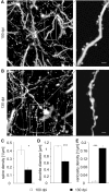
Figure 1.
Figure 1.
Typical spine and dendritic alterations of presymptomatic compared with symptomatic scrapie-infected mice. A, B, Representative images from two-photon in vivo imaging sessions of apical dendrites and adjacent spines of presymptomatic (100 dpi) and symptomatic (130 dpi) scrapie-infected Thy-1 YFP expressing mice. In contrast to the intact dendrites and unchanged spine density of presymptomatic mice, symptomatic mice exhibit spine loss and an increased number of dendritic swellings (varicosities). C, Mean spine density (1 per micrometer of dendrite length) is significantly reduced in symptomatic mice compared with presymptomatic mice (*p = 0.017). D, Symptomatic scrapie-infected mice exhibited a reduced mean dendrite diameter (in micrometers) compared with presymptomatic mice (***p = 9.9 × 10⁻¹⁴). E, Dendrites of presymptomatic mice display fewer varicosities per dendrite length compared with symptomatic mice (***p = 6.4 × 10⁻²³). Scale bars: A, B, 10 μm; insets, 2 μm.
Figure 2.
Figure 2.
Spines and dendritic varicosities are stable over 3 h throughout the presymptomatic and symptomatic phases of the disease. A, Repeated imaging of dendritic elements of scrapie-infected mice at 100 and 130 dpi. Note that spines (arrowheads) and varicosities (circles) are stable over the entire imaging period. B, Spine number, dendrite diameter, and number of varicosities do not change within 3 h of imaging (spines, p = 0.4; dendrite, p = 0.9; varicosities, p = 0.9). Scale bars, 2 μm.
Figure 3.
Figure 3.
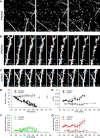
Kinetics of spine gain and loss of scrapie-infected and control mice from the presymptomatic to the symptomatic phase of prion disease. A, Low-magnification time-lapse images of a dendritic arbor part in the somatosensory cortex of a scrapie-infected YFP-H animal from the presymptomatic to terminal phase. B, Time series of a dendritic branch (from boxed region in A). Blue arrows exemplarily indicate persistent spines, red arrows point at lost spines, and green arrows specify gained spines. Note that persistent spines are continuously lost during the symptomatic phase of the disease. Spine loss is not compensated by the gain of spines. C, Time-lapse images of a dendritic branch of a control mouse imaged over a similar period (44 d) as scrapie-infected mice. Most of the spines are persistent (blue arrows), and spine loss and gain are balanced. D, Spine densities of scrapie-infected (filled circles) and control (open circles) mice. Lines correspond to individual animals and circles represent imaging days. Time-point values represent mean spine densities of 5–8 independent dendritic branches. Whereas the spine density of control mice remained constant, scrapie-infected animals exhibited a constant decrease of spine density from the presymptomatic to terminal phase of the disease. E, Mean daily spine turnover ratios of scrapie-infected (filled circles; n = 3 animals) and control mice (open circles; n = 3 animals). F, Average gained spine fraction per day of scrapie-infected and control mice (n = 3 animals each). During the presymptomatic phase of the disease, spine-gain values of diseased mice are comparable with control animals. Accompanied by the onset of the symptomatic phase of disease at 120 dpi, scrapie-infected animals exhibit an increased fraction of gained spines. G, Average lost spine fraction of scrapie-infected and control mice (n = 3 animals each). Already during the presymptomatic phase of the disease, scrapie-infected animals display a higher fraction of lost spines compared with controls. From 135 dpi on, highly increased spine loss occurs. Note that in scrapie-infected animals, a period of increased spine gain (120–135 dpi; F) precedes a period of increasing spine loss (135–150 dpi; G). Scale bars: A, 5 μm; B, C, 2 μm.
Figure 4.
Figure 4.
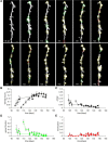
Structural plasticity of varicosities in scrapie-infected mice from the presymptomatic to the symptomatic phase of the disease. A, Representative time-lapse images of a dendritic branch of a scrapie-infected YFP-H mouse. Green circles represent gained, yellow circles indicate persistent, and red circles point at lost varicosities. Note that a large number of varicosities were gained during the presymptomatic phase of the disease until 120 dpi. Of the gained varicosities, 90 ± 2.7% persisted throughout the entire imaging period. Loss of varicosities was rarely observed. B, Density of varicosities from the beginning of the presymptomatic to the end of the symptomatic phase of the disease. Lines correspond to individual animals and symbols represent imaging days. Time-point values represent mean densities of varicosities of 5–8 independent dendritic branches. Varicosity densities of scrapie-infected animals exhibited a constant increase from the presymptomatic to the symptomatic phase of the disease, reaching a plateau from 130 dpi on. C, Mean daily varicosity turnover ratios of scrapie-infected mice. D, Average gained varicosity fraction of scrapie-infected mice. During the presymptomatic phase of disease, varicosity gain was greater than after the onset of the symptomatic phase at 120 dpi. E, Average lost varicosity fraction per day of scrapie-infected mice. Varicosity loss remained constantly low throughout the entire imaging period. Scale bar, 2.5 μm.
Figure 5.
Figure 5.
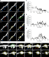
Dendritic varicosities predominantly gained at spines during the presymptomatic phase of prion disease subsequently loose adjacent spines during the symptomatic phase. A, Representative time series of a scrapie-infected YFP-H mouse. Persistent spines (blue arrowheads) were lost (red arrowheads) during the symptomatic phase of the disease. Varicosities (gained, green circles; persistent, yellow circles) mainly appeared at dendritic sites where spines protrude. Subsequently, a large number of spines was lost (red arrowheads) at gained or persistent varicosities. Rarely, new spines (green arrowheads) were gained at varicosities. B, High-magnification time-lapse images of dendritic spines. A varicosity was gained where spines protrude from the dendrite. Subsequently spines were lost at the varicosity that persisted until the end of the imaging period. C, Time-lapse of the average spine-gained density of varicosities in scrapie-infected mice. The density of spine-gained varicosities started at high values during the presymptomatic phase of the disease and decreased to nearly zero during the terminal phase (after 135 dpi). D, Time course of the average dendrite-gained density varicosities in scrapie-infected mice. The density of dendrite-gained varicosities initiated at small values during the presymptomatic phase of the disease and increased during the symptomatic phase (from 120 dpi). E, Time course of the mean varicosity-lost spine density of scrapie-infected YFP-H mice. Spine loss at varicosities is infrequent during the presymptomatic phase of disease. The density of spines lost at varicosities raised throughout the symptomatic phase of the disease, reaching a peak ∼130 dpi, and decreased until the end of imaging period. C–E, Each line represents an animal and symbols indicate imaging days. Scale bars: A, 2 μm; B, 1 μm.

References

    1. Akulinin VA, Semchenko VV, Stepanov SS, Belichenko PV. Structural changes in the dendritic spines of pyramidal neurons in layer III of the sensorimotor cortex of the rat cerebral cortex in the late post-ischemic period. Neurosci Behav Physiol. 2004;34:221–227. - PubMed
    1. Belichenko PV, Dahlstrom A. Studies on the 3-dimensional architecture of dendritic spines and varicosities in human cortex by confocal laser scanning microscopy and Lucifer yellow microinjections. J Neurosci Methods. 1995;57:55–61. - PubMed
    1. Belichenko PV, Brown D, Jeffrey M, Fraser JR. Dendritic and synaptic alterations of hippocampal pyramidal neurones in scrapie-infected mice. Neuropathol Appl Neurobiol. 2000;26:143–149. - PubMed
    1. Bouzamondo-Bernstein E, Hopkins SD, Spilman P, Uyehara-Lock J, Deering C, Safar J, Prusiner SB, Ralston HJ, III, DeArmond SJ. The neurodegeneration sequence in prion diseases: evidence from functional, morphological and ultrastructural studies of the GABAergic system. J Neuropathol Exp Neurol. 2004;63:882–899. - PubMed
    1. Brown D, Belichenko P, Sales J, Jeffrey M, Fraser JR. Early loss of dendritic spines in murine scrapie revealed by confocal analysis. NeuroReport. 2001;12:179–183. - PubMed

Publication types

LinkOut - more resources