Comparative Study
. 2007 Jun 6;27(23):6261-7.
doi: 10.1523/JNEUROSCI.5646-06.2007.

A novel connection between rods and ON cone bipolar cells revealed by ectopic metabotropic glutamate receptor 7 (mGluR7) in mGluR6-deficient mouse retinas

Affiliations
Comparative Study

A novel connection between rods and ON cone bipolar cells revealed by ectopic metabotropic glutamate receptor 7 (mGluR7) in mGluR6-deficient mouse retinas

Yoshihiko Tsukamoto et al. J Neurosci. .

Abstract

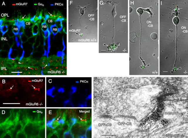
Since the discovery of direct chemical synapses between rod photoreceptor and OFF cone bipolar cells in mouse retinas, whether the ON cone bipolar cell also receive direct chemical input from rod has been a pending question. In finding that metabotropic glutamate receptor 7 (mGluR7) was uniquely expressed in dendrites of ON cone bipolar cells in the mGluR6-deficient mouse retina, we used this ectopic mGluR7 immunoreactivity as a specific marker for the ON cone bipolar to search for its rod connection. Here, we show that a certain type of ON cone bipolar cell forms ribbon-associated synapses not only with cones, but also rods. This finding was verified in the wild-type mouse retina by three-dimensional reconstruction of bipolar cells from serial electron micrographs. These ON cone bipolars were further identified as corresponding to type 7 of mouse bipolar cell described by Ghosh et al. (2004) and also to the green fluorescent protein (GFP)-labeled type 7 bipolars in the alpha-gustducin-GFP transgenic mouse. Our findings suggest that, in mice, rod signals bifurcate into a third ON and OFF pathway in addition to the two known routes to cone bipolar cells: (1) via rod chemical synapse --> rod bipolar --> AII amacrine --> ON and OFF cone bipolar cells; (2) via rod-cone gap junction --> cone chemical synapse --> ON and OFF cone bipolar cells; and (3) via rod chemical synapse --> ON and OFF cone bipolar cells. This third novel pathway is thought to transmit fast and moderately light-sensitive rod signals, functioning to smooth out the intensity changes at the scotopic-mesopic interface.


Figures

Figure 1.
Figure 1.
Immunocytochemistry of cone bipolar (CB) cells using the anti-mGluR7 antibody. A–E, The mGluR6-deficient mouse retina is triply stained for mGluR7 in red, for Gαo in green (labeling both ON cone and rod bipolar cells), and for PKCα in blue (labeling only rod bipolar cells). Intermittent red patches in the OPL coincide with green areas (arrows). Numerous red patches (arrowheads) are also scattered in the IPL. Green areas overlap blue areas, but red patches never coincide with any blue areas. F–I, Dissociated OFF (F, G) and ON (H, I) cone bipolar cells are immunolabeled for mGluR7 (green) and observed by differential interference contrast. The dendrites are mGluR7-immunoreactive (arrow) only for the ON cone bipolar cell of mGluR6-deficient (−/−) mouse (I). The axon terminals are always mGluR7-immunoreactive (arrowheads) for both OFF and ON bipolar cells regardless of the sufficiency (+/+) or deficiency (−/−) of mGluR6. The dissociated cone bipolar cells appear to correspond to the types of mouse bipolar cells described by Ghosh et al. (2004) as follows: F, type 2; G, type 3; H, type 6 or 7; and I, type 6 or 7. J, Electron micrograph of a cone synaptic terminal in the mGluR6−/− mouse retina immunolabeled for mGluR7. One immunoreactive dendritic process invaginates the cone pedicle up to a synaptic site facing the cone ribbon. The adjacent smaller immunoreactive process was confirmed to invaginate the cone pedicle in other sections. These two processes are morphologically identified as dendrites of the ON cone bipolar (ON-CB) cells. At the neighboring basal surface of the pedicle, there are two flat synaptic contacts. Their postsynaptic processes are morphologically identified as the dendrites of OFF cone bipolar (OFF-CB) cells, which show no mGluR7 immunoreactivity. Scale bars: A, B, G, I, 5 μm; J, 200 nm.
Figure 2.
Figure 2.
A novel synaptic junction between rod photoreceptor and ON cone bipolar cell in mGluR6-deficient mouse. A–D, Rod and cone contacts with mGluR7-labeled ON cone bipolar dendrites (red) in the mGluR6-deficient mouse retina. A ribbon (green) in a rod spherule (RS) is located above a cone pedicle (CP) and shaped like a horseshoe. Ribbons (green) in CPs are relatively small and aligned as clusters. B, A dendritic tip labeled with mGluR7 immunoreactivity (white arrow) makes contact with the ribbon (white arrowhead) in the RS. A larger dendrite labeled with mGluR7 immunoreactivity (yellow arrow) branches out in apposition to ribbons (yellow arrowheads) in the CP. E–H, Electron micrographs of serial sections from the mGluR6−/− mouse retina immunolabeled for mGluR7. One process is mGluR7-immunoreactive (arrow) and invaginates the rod spherule up to a synaptic ribbon (arrowhead). Nu, The nucleus of a rod photoreceptor cell.
Figure 3.
Figure 3.
Cone and rod mixed input type of bipolar cell in wild-type mouse. A, B, Two adjacent serial electron micrographs of the synaptic terminal of a rod photoreceptor in a wild-type mouse retina, where two invaginating bipolar processes form similar synapses in association with a ribbon (arrowhead). One process comes from a rod bipolar cell (RB) and the other from an ON cone bipolar cell (CB). H, Horizontal cell processes. C, Type B7 ON cone bipolar dendrites have ribbon synapses with rods as well as cones (the reconstructed B7 cell in D at higher magnification). D, Reconstruction, from the same serial electron micrographs as in A and B, of mouse ON cone bipolar cells typed B5, B6, B7, and B8. The depth of the axon terminal in the IPL is 57.8 ± 6.1% (n = 4) for type B5, 89.0 ± 4.5% (n = 3) for type B6, 73.5 ± 4.4% (n = 3) for type B7, and 90.2% (n = 1) for type B8. Asterisks for cell B8 indicate the end points of the series of electron micrographs.
Figure 4.
Figure 4.
A rod contact and cone contacts with GFP-labeled type 7 bipolar dendrites (green) in the (mGluR6+/+) GUS-GFP mouse retina. A–C, A CtBP2-labeled ribbon (red) in a rod spherule (RS) lies singly and spans three optical sections (∼1.3 μm in total thickness). A single dendritic process (white arrow) ascends toward the RS and appears to make contact with the ribbon (white arrowhead). B–D, CtBP2-labeled ribbons (red) in cone pedicles (CPs) are relatively small and aligned in clusters. Two dendrites (yellow arrows) branch out in close apposition to ribbons (yellow arrowheads) in CPs.

References

    1. Brandstätter JH, Koulen P, Kuhn R, van der Putten H, Wässle H. Compartmental localization of a metabotropic glutamate receptor (mGluR7): two different active sites at a retinal synapse. J Neurosci. 1996;16:4749–4756. - PMC - PubMed
    1. DeVries SH, Li W, Saszik S. Parallel processing in two transmitter microenvironments at the cone photoreceptor synapse. Neuron. 2006;50:735–748. - PubMed
    1. Dhingra A, Lyubarsky A, Jiang M, Pugh EN, Jr, Birnbaumer L, Sterling P, Vardi N. The light response of ON bipolar neurons requires Gαo. J Neurosci. 2000;20:9053–9058. - PMC - PubMed
    1. Dowling JE. Cambridge, MA: Harvard UP; 1987. The retina.
    1. Ghosh KK, Bujan S, Haverkamp S, Feigenspan A, Wässle H. Types of bipolar cells in the mouse retina. J Comp Neurol. 2004;469:70–82. - PubMed

Publication types

MeSH terms

Substances

LinkOut - more resources