Review
. 2007 Jun;71(2):282-94.
doi: 10.1128/MMBR.00037-06.

A biochemical guide to yeast adhesins: glycoproteins for social and antisocial occasions

Affiliations
Review

A biochemical guide to yeast adhesins: glycoproteins for social and antisocial occasions

Anne M Dranginis et al. Microbiol Mol Biol Rev. 2007 Jun.

Abstract

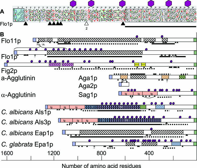
Fungi are nonmotile eukaryotes that rely on their adhesins for selective interaction with the environment and with other fungal cells. Glycosylphosphatidylinositol (GPI)-cross-linked adhesins have essential roles in mating, colony morphology, host-pathogen interactions, and biofilm formation. We review the structure and binding properties of cell wall-bound adhesins of ascomycetous yeasts and relate them to their effects on cellular interactions, with particular emphasis on the agglutinins and flocculins of Saccharomyces and the Als proteins of Candida. These glycoproteins share common structural motifs tailored to surface activity and biological function. After being secreted to the outer face of the plasma membrane, they are covalently anchored in the wall through modified GPI anchors, with their binding domains elevated beyond the wall surface on highly glycosylated extended stalks. N-terminal globular domains bind peptide or sugar ligands, with between millimolar and nanomolar affinities. These affinities and the high density of adhesins and ligands at the cell surface determine microscopic and macroscopic characteristics of cell-cell associations. Central domains often include Thr-rich tandemly repeated sequences that are highly glycosylated. These domains potentiate cell-to-cell binding, but the molecular mechanism of such an association is not yet clear. These repeats also mediate recombination between repeats and between genes. The high levels of recombination and epigenetic regulation are sources of variation which enable the population to continually exploit new niches and resources.


Figures

FIG. 1.
FIG. 1.
Molecular features of representative yeast adhesins. These features are based on HCA plots of the adhesins. (A) HCA plot of the N-terminal 440 residues of Flo1p. HCA draws each open reading frame as a helical projection, which is vertically repeated. Individual amino acids are plotted and colored red for acidic, blue for basic, and green for hydrophobic, with hydrophobic patches bounded by black lines. Thr residues are drawn as hollow squares, Ser as dark-centered squares, Gly as diamonds, and Pro as red stars. Cys residues are marked with triangles below the HCA plot, and N-glycosylation sites are marked with maroon hexagons. Transparent boxes designate the N-terminal secretion signal (light blue) and the beginning of a tandem repeat region (open box). The line below the right-hand end shows that the Thr content of the repeat region is >25%. (B) Summary of HCAs of representative yeast adhesins, aligned at the C termini, where they are linked to cell wall polysaccharide through the GPI anchor remnant. N-terminal secretion signals are light blue, and GPI addition signals are green. Repeated sequences are boxed. Diagonal stripes in white boxes indicate tandem repeats that are not homologous to other repeats in the illustrated proteins. Where similar sequences recur, they are boxed and tinted in the same color. Potential N-glycosylation sites are shown as maroon hexagons above each open reading frame. Cys residues are shown as triangles below each open reading frame and are linked where the disulfide bonds have been mapped. The content of Thr is denoted by bars below each open reading frame, which are dotted where the Thr content exceeds 20% and solid where it exceeds 25%. (The frequency in the entire S. cerevisiae genome is 6%.)
FIG. 2.
FIG. 2.
Adhesin concentration in vivo. A scale drawing shows complementary 100,000-Da adhesins displayed on cell walls of two apposing yeast cells at the “moderate” surface concentration of about 2.5 × 10⁴ molecules per cell, corresponding to a cell surface concentration of ∼4 × 10⁻⁴ M. This surface concentration is approximately equivalent to that in a solution with 40 mg/ml of each of the adhesins, which is 100-fold greater than the usual concentrations in biochemical experiments with these proteins. The cell diameters are 4 μm, and a 1-μm by 1-μm section of each cell wall is shown as a curved tan sector with internal structure omitted. The lengths of proteins (∼100 nm) are drawn to scale, but the thickness is exaggerated to improve visibility.
FIG. 3.
FIG. 3.
Several phenotypes associated with the adhesin Flo11p. (A) Flocculation of S. cerevisiae var. diastaticus is Flo11 dependent. Yeast cultures of equal cell densities were vortexed vigorously and photographed at the indicated time intervals after mixing. Left tubes, wild-type haploid cells; right tubes, isogenic yeast with FLO11 deletion. (B) S. cerevisiae strain Σ1278b form Flo11-dependent biofilms. A single colony of yeast is shown growing on semisolid medium in a standard 100-mm petri dish (101). The biofilm with its characteristic floral structure requires Flo11 for its formation. (C) Diploid Σ1278b strains form branched chains of cells called pseudohyphae in response to nitrogen starvation. Formation of these chains requires Flo11p (81).

Similar articles

Cited by

References

    1. Andrews, J., and R. B. Gilliland. 1952. Super-attenuation of beer: a study of three organisms capable of causing abnormal attenuation. J. Inst. Brewing 58:189-196.
    1. Batlle, M., A. Lu, D. A. Green, Y. Xue, and J. P. Hirsch. 2003. Krh1p and Krh2p act downstream of the Gpa2p G(alpha) subunit to negatively regulate haploid invasive growth. J. Cell Sci. 116:701-710. - PubMed
    1. Bayly, J. C., L. M. Douglas, I. S. Pretorius, F. F. Bauer, and A. M. Dranginis. 2005. Characteristics of Flo11-dependent flocculation in Saccharomyces cerevisiae. FEMS Yeast Res. 5:1151-1156. - PubMed
    1. Bidard, F., B. Blondin, S. Dequin, F. Vezinhet, and P. Barre. 1994. Cloning and analysis of a FLO5 flocculation gene from S. cerevisiae. Curr. Genet. 25:196-201. - PubMed
    1. Bidard, F., M. Bony, B. Blondin, S. Dequin, and P. Barre. 1995. The Saccharomyces cerevisiae FLO1 flocculation gene encodes for a cell surface protein. Yeast 11:809-822. - PubMed

Publication types

MeSH terms

LinkOut - more resources