Review
. 2007 Jun;71(2):295-347.
doi: 10.1128/MMBR.00040-06.

Sponge-associated microorganisms: evolution, ecology, and biotechnological potential

Affiliations
Review

Sponge-associated microorganisms: evolution, ecology, and biotechnological potential

Michael W Taylor et al. Microbiol Mol Biol Rev. 2007 Jun.

Abstract

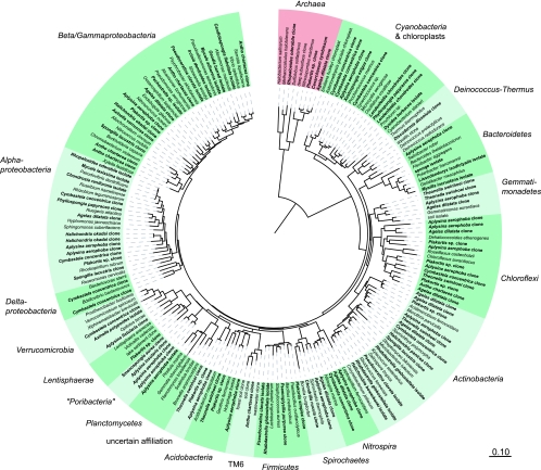
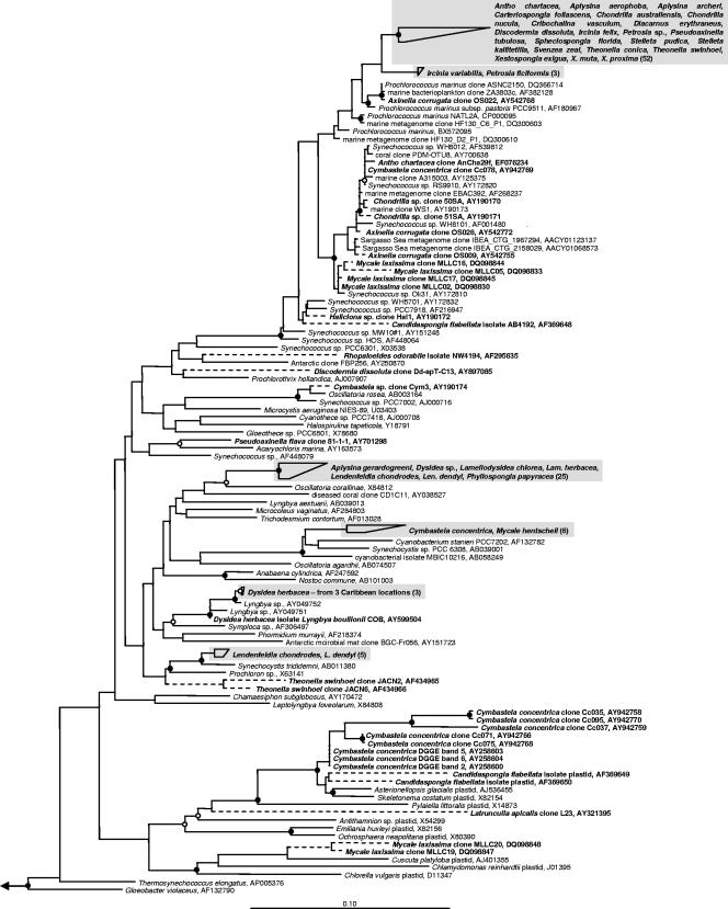
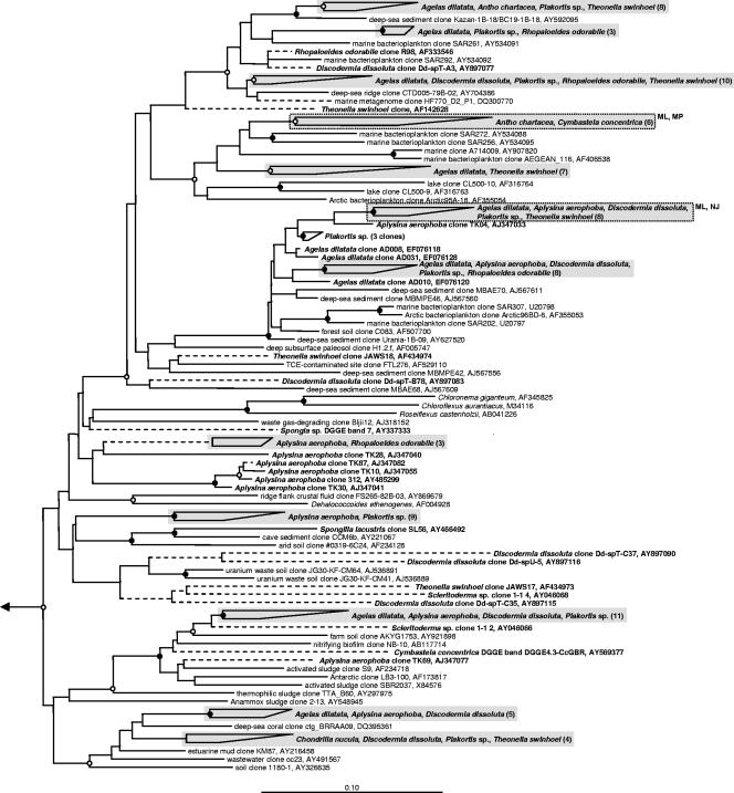
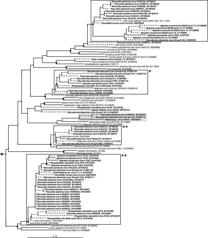
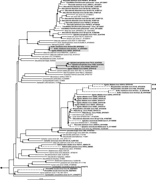
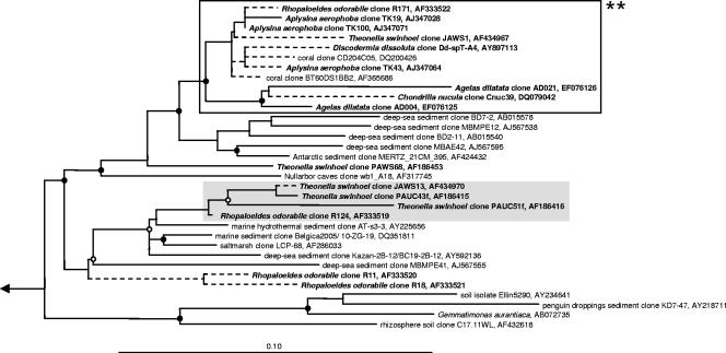
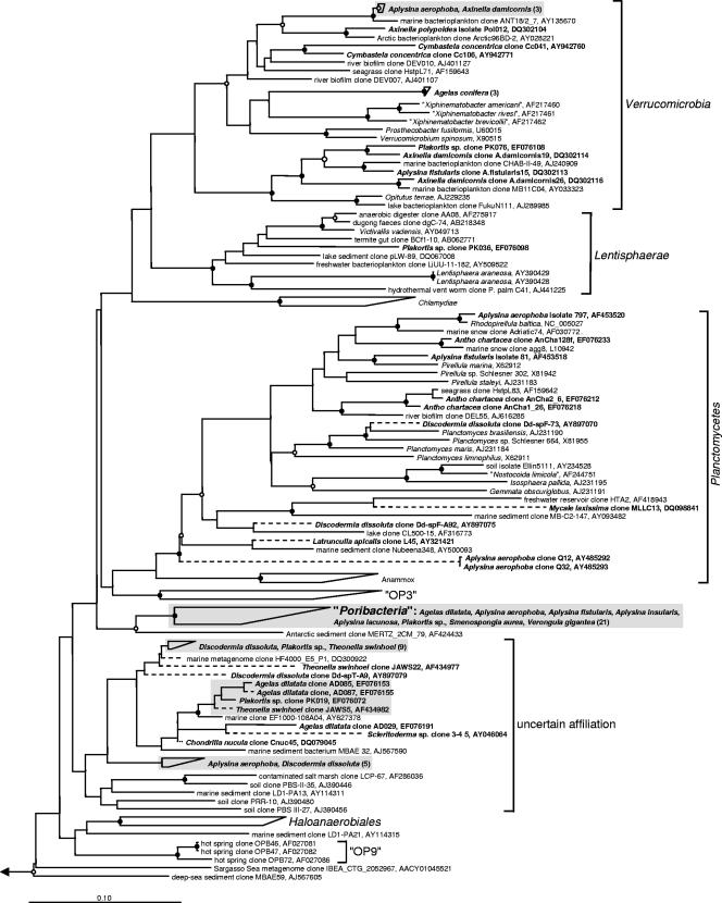
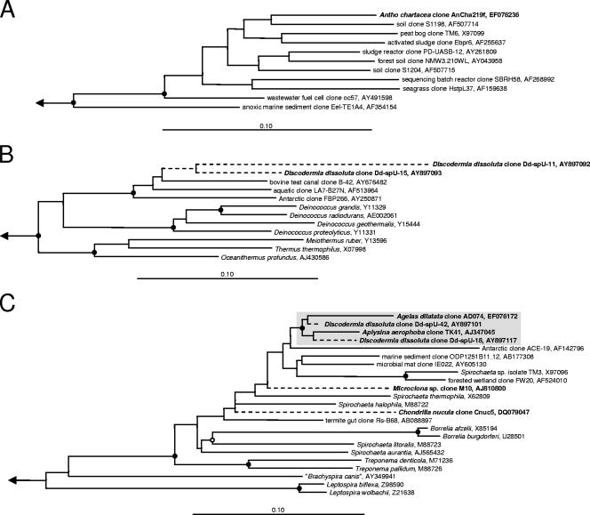
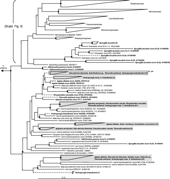
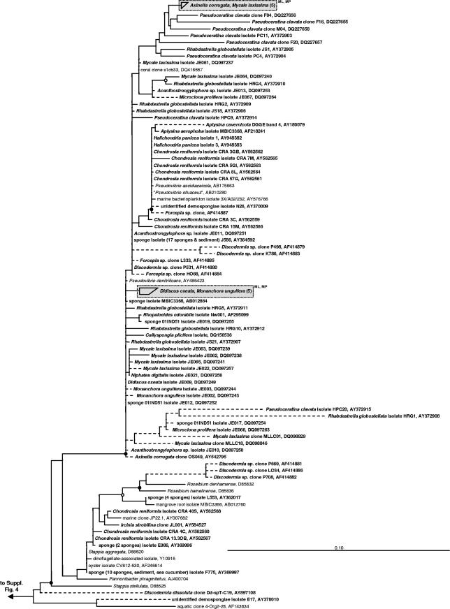
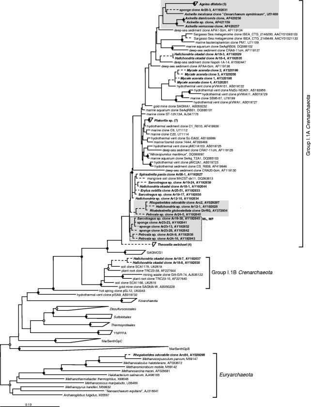
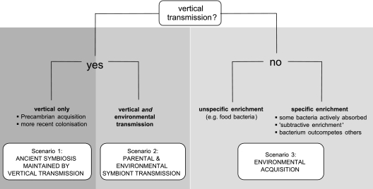
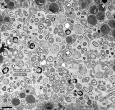
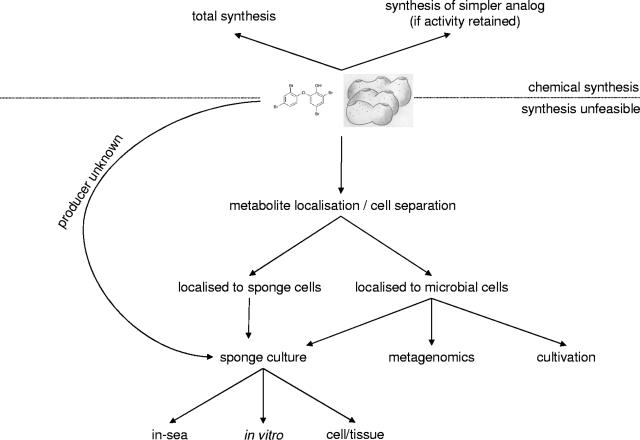
Marine sponges often contain diverse and abundant microbial communities, including bacteria, archaea, microalgae, and fungi. In some cases, these microbial associates comprise as much as 40% of the sponge volume and can contribute significantly to host metabolism (e.g., via photosynthesis or nitrogen fixation). We review in detail the diversity of microbes associated with sponges, including extensive 16S rRNA-based phylogenetic analyses which support the previously suggested existence of a sponge-specific microbiota. These analyses provide a suitable vantage point from which to consider the potential evolutionary and ecological ramifications of these widespread, sponge-specific microorganisms. Subsequently, we examine the ecology of sponge-microbe associations, including the establishment and maintenance of these sometimes intimate partnerships, the varied nature of the interactions (ranging from mutualism to host-pathogen relationships), and the broad-scale patterns of symbiont distribution. The ecological and evolutionary importance of sponge-microbe associations is mirrored by their enormous biotechnological potential: marine sponges are among the animal kingdom's most prolific producers of bioactive metabolites, and in at least some cases, the compounds are of microbial rather than sponge origin. We review the status of this important field, outlining the various approaches (e.g., cultivation, cell separation, and metagenomics) which have been employed to access the chemical wealth of sponge-microbe associations.


Figures

FIG. 1.
FIG. 1.
Increasing research interest in marine sponge-microorganism associations. (A) Number of publications retrieved from the ISI Web of Science database by using the following search string: (sponge* or porifera* or demospong* or sclerospong* or hexactinellid*) and (bacteri* or prokaryot* or microbe* or microbial or microorganism* or cyanobacteri* or archaeon or archaea* or crenarchaeo* or fung* or diatom* or dinoflagellate* or zooxanthella*) not (surgery or surgical). (B) Number of sponge-derived 16S rRNA gene sequences deposited in GenBank per year. The 2006 value includes the 184 sequences submitted to GenBank from this article. The search string used to recover sequences was as follows: (sponge* or porifera*) and (16S* or ssu* or rRNA*) not (18S* or lsu* or large subunit or mitochondri* or 23S* or 5S* or 5.8S* or 28S* or crab* or alga* or mussel* or bivalv* or crustacea*).
FIG. 2.
FIG. 2.
Schematic representation of a sponge. Arrows indicate the direction of water flow through the sponge. (Adapted from reference with permission of Brooks/Cole, a division of Thomson Learning.)
FIG. 3.
FIG. 3.
Sponges of diverse size, shape, and color. The encrusting sponge Tedania digitata (left), the branching sponge Axinella cannabina (center), and the giant barrel sponge Xestospongia testudinaria (right) are shown. The last two images were kindly provided by Armin Svoboda (Ruhr-Universität, Bochum, Germany).
FIG. 4.
FIG. 4.
16S rRNA-based phylogeny showing representatives of all bacterial and archaeal phyla from which sponge-derived sequences have been obtained. Sponge-derived sequences are shown in bold, with additional reference sequences also included. The displayed tree is based on a maximum likelihood analysis. Bar, 10% sequence divergence.
FIG. 5.
FIG. 5.
16S rRNA-based phylogeny of sponge-associated cyanobacteria and chloroplasts. Sponge-derived sequences are shown in bold. The displayed tree is a maximum likelihood tree constructed based on long (≥1,000 nucleotides) sequences only. Shorter sequences were added using the parsimony interactive tool in ARB and are indicated by dashed lines. Shaded boxes represent sponge-specific monophyletic clusters, as defined by Hentschel et al. (146), i.e., a group of at least three sponge-derived 16S rRNA gene sequences which (i) are more similar to each other than to sequences from other, nonsponge sources, (ii) are found in at least two host sponge species and/or in the same host species but from different geographic locations, and (iii) cluster together irrespective of the phylogeny inference method used (all clusters shown here also occurred in neighbor-joining and maximum parsimony analyses). Names outside wedges of grouped sequences represent the sponges from which the relevant sequences were derived; the number in parentheses indicates the number of sequences in that wedge. Filled circles indicate bootstrap support (maximum parsimony, with 100 resamplings) of ≥90%, and open circles represent ≥75% support. The outgroup (not shown) consisted of a range of sequences representing several other bacterial phyla. Bar, 10% sequence divergence.
FIG. 6.
FIG. 6.
16S rRNA-based phylogeny of sponge-associated Chloroflexi organisms. Details are the same as those provided for Fig. 5, with the following additions. Shaded boxes contained within dotted lines represent sponge-specific clusters supported by only two tree construction methods (ML, maximum likelihood; MP, maximum parsimony; and NJ, neighbor joining), and new sequences from our laboratory have the prefix “AD” (for the sponge Agelas dilatata), “AnCha” (Antho chartacea), or “PK” (Plakortis sp.).
FIG. 7.
FIG. 7.
16S rRNA-based phylogeny of sponge-associated Acidobacteria organisms. Details are the same as those provided for Fig. 5 and 6, with the following two additions. Open boxes represent monophyletic clusters containing sponge-derived sequences and a single, nonsponge origin sequence, and open boxes with asterisks outside them signify clusters containing only sponge- and coral-derived sequences (the number of asterisks corresponds to the number of coral-derived sequences within the cluster).
FIG. 8.
FIG. 8.
16S rRNA-based phylogeny of sponge-associated Deltaproteobacteria organisms. Details are the same as those provided for Fig. 5 to 7.
FIG. 9.
FIG. 9.
16S rRNA-based phylogeny of sponge-associated Gemmatimonadetes organisms. Details are the same as those provided for Fig. 5 to 7.
FIG. 10.
FIG. 10.
16S rRNA-based phylogeny of sponge-associated Nitrospira organisms. Details are the same as those provided for Fig. 5 to 7.
FIG. 11.
FIG. 11.
16S rRNA-based phylogeny of sponge-associated Verrucomicrobia, Planctomycetes, Lentisphaerae, and “Poribacteria” organisms and of a lineage of uncertain affiliation. These and associated lineages comprising the PVC superphylum (446) are shown. Details are the same as those provided for Fig. 5 to 7.
FIG. 12.
FIG. 12.
16S rRNA-based phylogeny of sponge-associated members of the candidate phylum TM6 (A), Deinococcus-Thermus organisms (B), and Spirochaetes organisms (C). Details are the same as those provided for Fig. 5 to 7. (B) In our analyses, the position of clone Dd-spU-11 (from the sponge Discodermia dissoluta) was not stable, and we are not certain of its phylogenetic affiliation.
FIG. 13.
FIG. 13.
16S rRNA-based phylogeny of sponge-associated Actinobacteria organisms belonging to the family Acidimicrobiaceae. Other sponge-derived actinobacteria are shown in Fig. S3 in the supplemental material. Details are the same as those provided for Fig. 5 to 7.
FIG. 14.
FIG. 14.
16S rRNA-based phylogeny of sponge-associated Alphaproteobacteria organisms affiliated with the genus Pseudovibrio and its relatives. Other sponge-derived alphaproteobacteria are shown in Fig. S4 and S5 in the supplemental material. Details are the same as those provided for Fig. 5 to 7.
FIG. 15.
FIG. 15.
16S rRNA-based phylogeny of sponge-associated archaeal organisms. Details are the same as those provided for Fig. 5 to 7.
FIG. 16.
FIG. 16.
Summary of various evolutionary scenarios for sponge-microorganism associations.
FIG. 17.
FIG. 17.
Vertical transmission of microbial symbionts by a marine sponge. A transmission electron micrograph of a Chondrilla australiensis larva is shown, indicating a diverse range of bacterial morphotypes. Bar = 1 μm. (Modified from reference with permission of the publisher.)
FIG. 18.
FIG. 18.
Current state of knowledge about the nitrogen cycle in sponges. Thick arrows signify those processes which have been demonstrated in sponges; references (given in parentheses) pertain to either the process or the implicated microorganisms. PON, particulate organic nitrogen.
FIG. 19.
FIG. 19.
Effect of a bacterial pathogen on a marine sponge. Transmission electron micrographs of Rhopaloeides odorabile tissue are shown, displaying (A) the diversity of bacterial morphotypes in healthy tissue, (B) a sponge experimentally infected with the alphaproteobacterial pathogen strain NW4327, and (C) consequent necrosis of the sponge tissue. Bar = 500 nm. (Reprinted from reference with permission of the publisher.)
FIG. 20.
FIG. 20.
Global distributions of selected monophyletic, sponge-specific clusters. Symbols refer to collection locations for representatives of the “Ca. Synechococcus spongiarum” (Cyanobacteria) (circles), Actinobacteria (triangles), and Acidobacteria (stars) clusters. In the last cluster, coral-derived sequences from the Mediterranean are also present.
FIG. 21.
FIG. 21.
Chemical structures of jaspamide (left), from Jaspis sp. sponges, and chondramide D (right), from the deltaproteobacterium Chondromyces crocatus. Note the remarkable structural similarities between the compounds.
FIG. 22.
FIG. 22.
Chemical structure of halichondrin B.
FIG. 23.
FIG. 23.
Chemical structure of peloruside A.
FIG. 24.
FIG. 24.
Approaches for obtaining bioactive metabolites from marine sponges.
FIG. 25.
FIG. 25.
In-sea aquaculture of the Great Barrier Reef sponge Rhopaloeides odorabile. (Image courtesy of Rocky de Nys [James Cook University, Australia], reproduced with permission.)

Similar articles

Cited by

References

    1. Adamczeski, M., A. R. Reed, and P. Crews. 1995. New and known diketopiperazines from the Caribbean sponge, Calyx cf. podatypa. J. Nat. Prod. 58:201-208. - PubMed
    1. Adamczyk, J., M. Hesselsoe, N. Iversen, M. Horn, A. Lehner, P. H. Nielsen, M. Schloter, P. Roslev, and M. Wagner. 2003. The isotope array, a new tool that employs substrate-mediated labeling of rRNA for determination of microbial community structure and function. Appl. Environ. Microbiol. 69:6875-6887. - PMC - PubMed
    1. Ahn, Y. B., S. K. Rhee, D. E. Fennell, L. J. Kerkhof, U. Hentschel, and M. M. Haggblom. 2003. Reductive dehalogenation of brominated phenolic compounds by microorganisms associated with the marine sponge Aplysina aerophoba. Appl. Environ. Microbiol. 69:4159-4166. - PMC - PubMed
    1. Aicher, T. D., K. R. Buszek, F. G. Fang, C. J. Forsyth, S. H. Jung, Y. Kishi, M. C. Matelich, P. M. Scola, D. M. Spero, and S. K. Yoon. 1992. Total synthesis of halichondrin B and norhalichondrin B. J. Am. Chem. Soc. 114:3162-3164.
    1. Aizenberg, J., V. C. Sundar, A. D. Yablon, J. C. Weaver, and G. Chen. 2004. Biological glass fibers: correlation between optical and structural properties. Proc. Natl. Acad. Sci. USA 101:3358-3363. - PMC - PubMed

Publication types

LinkOut - more resources