

doi: 10.1107/S1744309107024487.
Epub 2007 May 31.
Crystallization and preliminary X-ray analysis of PH1010 from Pyrococcus horikoshii OT3, a member of the archaeal DUF54 family of proteins
Affiliations
- PMID: 17554180
- PMCID: PMC2335084
- DOI: 10.1107/S1744309107024487
Item in Clipboard
Crystallization and preliminary X-ray analysis of PH1010 from Pyrococcus horikoshii OT3, a member of the archaeal DUF54 family of proteins
Acta Crystallogr Sect F Struct Biol Cryst Commun.
.
Abstract
PH1010 from Pyrococcus horikoshii OT3, a member of the archaeal DUF54 family of proteins, was expressed, purified and crystallized. Crystallization was performed by the sitting-drop vapour-diffusion method using PEG 3350 as the precipitant. The crystal diffracted X-rays to 1.90 A resolution using a synchrotron-radiation source. The space group of the crystal was determined to be P2(1)2(1)2(1), with unit-cell parameters a = 46.9, b = 49.5, c = 132.7 A. The crystal contained two PH1010 molecules in the asymmetric unit (V(M) = 2.4 A(3) Da(-1)) and had a solvent content of 48%.
Figures
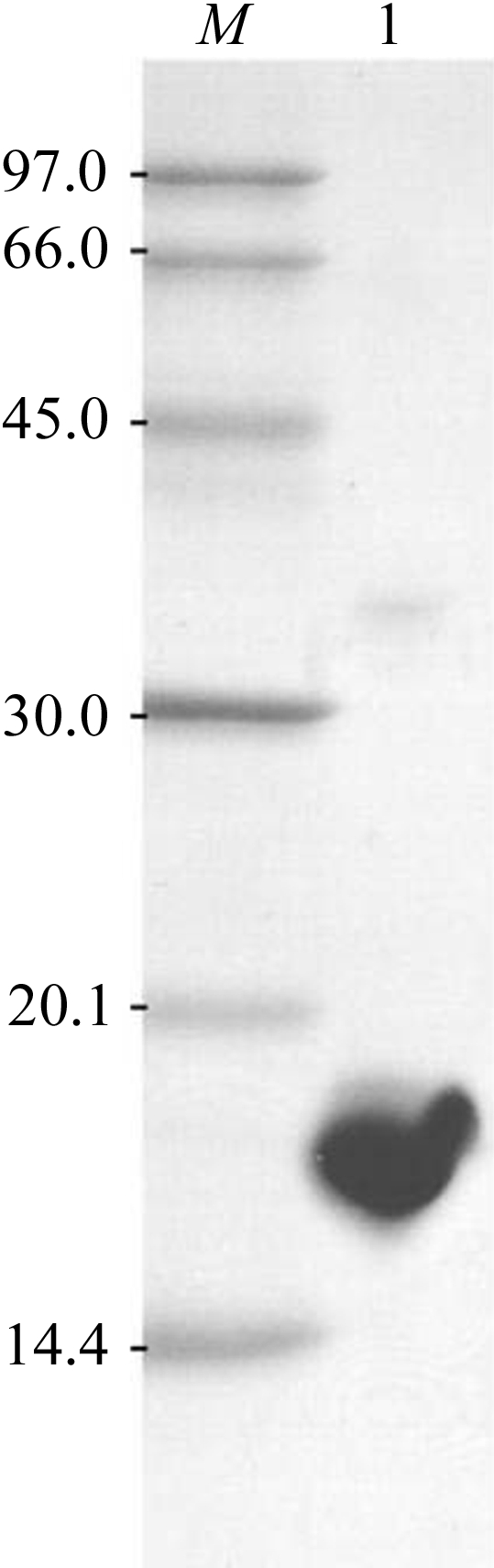
SDS–PAGE of the purified PH1010. Lane M, molecular-weight markers (kDa). Lane 1, PH1010.

Images of typical PH1010 crystals. (a) Crystals of the native protein of approximate dimensions 0.25 × 0.1 × 0.05 mm. (b) Crystals of the SeMet derivative of approximate dimensions 0.25 × 0.1 × 0.05 mm.

An X-ray diffraction image from a typical crystal of native protein. Diffraction data are detectable to 1.90 Å. The edge of the detector corresponds to a resolution of 1.90 Å.
References
-
- Bricogne, G., Vonrhein, C., Flensburg, C., Schiltz, M. & Paciorek, W. (2003). Acta Cryst. D59, 2023–2030. - PubMed
-
- Cohen, G. N., Barbe, V., Flament, D., Galperin, M., Heilig, R., Lecompte, O., Poch, O., Prieur, D., Querellou, J., Ripp, R., Thierry, J. C., Van der Oost, J., Weissenbach, J., Zivanovic, Y. & Forterre, P. (2003). Mol. Microbiol.47, 1495–1512. - PubMed
-
- Kawarabayasi, Y. et al. (1998). DNA Res.5, 55–76. - PubMed
Publication types
MeSH terms
Substances
LinkOut - more resources
Full Text Sources
