

Evaluating the degree of conformity of papillary carcinoma and follicular carcinoma to the reported ultrasonographic findings of malignant thyroid tumor
- PMID: 17554185
- PMCID: PMC2627417
- DOI: 10.3348/kjr.2007.8.3.192
Evaluating the degree of conformity of papillary carcinoma and follicular carcinoma to the reported ultrasonographic findings of malignant thyroid tumor
Abstract
Objective: We wanted to evaluate the degree of conformity of papillary carcinoma and follicular carcinoma to the reported ultrasonographic findings of malignant thyroid tumor.
Materials and methods: Between January 2003 and December 2004, fine needle aspiration biopsy was performed in 1,036 patients with palpable and nonpalpable thyroid lesions. We retrospectively reviewed the ultrasonographic findings of patients with papillary carcinomas (n = 127) and follicular carcinomas (n = 23) that were proven by operation or fine needle aspiration biopsy. We analyzed the ultrasonographic findings of these nodules based on the reported ultrasonographic findings of malignant thyroid tumor: hypoechogenicity, a taller than wide orientation, a microlobulated or irregular margin, a thick hypoechoic rim (halo sign), microcalcification and cystic change.
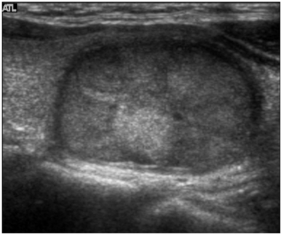
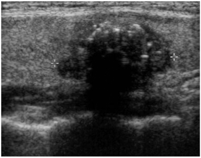
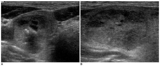
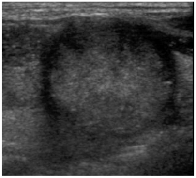
Results: The echogenicity was hypoechoic in 72.4% (92/127) of the papillary carcinomas, but it was isoechoic in 65.2% (15/23) of the follicular carcinomas (p < 0.001). The nodule shape was tall or round in 74.1% of the papillary carcinomas, but it was flat in 72.7% of the follicular carcinomas (p < 0.001). The tumor margin was microlobulated or irregular in 92.9% of the papillary carcinomas and in 60.9% of the follicular carcinomas (p < 0.001). A hypoechoic rim was seen in 26% of the papillary carcinomas (thin rim: 13.4%, thick rim: 12.6%) and in 86.6% of the follicular carcinomas (thin rim: 39.1%, thick rim: 47.8%, p < 0.001). Microcalcifications were demonstrated in 33.9% of the papillary carcinomas and in none of the cases of follicular carcinoma (p < 0.001). A solid mass without cystic change were seen in 98.4% of the papillary carcinomas and in 82.6% of the follicular carcinomas (p < 0.001).
Conclusion: The previously reported ultrasonography findings of malignant thyroid tumor are in conformity with most of the papillary carcinomas, but not with follicular carcinomas. The current ultrasonographic features for thyroid malignancy should be cautiously applied as the indication for needle aspiration biopsy so that follicular carcinomas are not missed by too narrow and strict biopsy criteria.
Figures
References
-
- Frates MC, Benson CB, Charbouneau JW, Cibas ES, Clark OH, Coleman BG, et al. Management of thyroid nodules detected at US: Society of Radiologists in Ultrasound consensus conference statement. Radiology. 2005;237:794–800. - PubMed
-
- Kim EK, Park CS, Chung WY, Oh KK, Kim DI, Lee JT, et al. New sonographic criteria for recommending fine-needle aspiration biopsy of nonpalpable solid nodules of the thyroid. AJR Am J Roentgenol. 2002;178:687–691. - PubMed
-
- Frates MC, Benson CB, Doubilet PM, Cibas ES, Marqusee E. Can color Doppler sonography aid in prediction of malignancy of thyroid nodules? J Ultrasound Med. 2003;22:127–131. - PubMed
-
- Papini E, Guglielmi R, Bianchini A, Crescenzi A, Taccogna S, Nardi F, et al. Risk of malignancy in nonpalpable thyroid nodules: predictive value of ultrasound and color-Doppler features. J Clin Endocrinol Metab. 2002;87:1941–1946. - PubMed
-
- Chan BK, Desser TS, McDougall IR, Weigel RJ, Jeffrey RB., Jr Common and uncommon sonographic features of papillary thyroid carcinoma. J Ultrasound Med. 2003;22:1083–1090. - PubMed
MeSH terms
LinkOut - more resources
Full Text Sources
Medical
