

Sonographically-guided 14-gauge core needle biopsy for papillary lesions of the breast
- PMID: 17554187
- PMCID: PMC2627411
- DOI: 10.3348/kjr.2007.8.3.206
Sonographically-guided 14-gauge core needle biopsy for papillary lesions of the breast
Abstract
Objective: We wanted to assess the need for surgical excising papillary lesions of the breast that were diagnosed upon sonographically guided 14-gauge core needle biopsy.
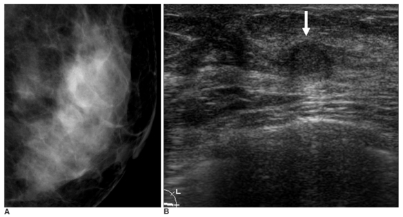
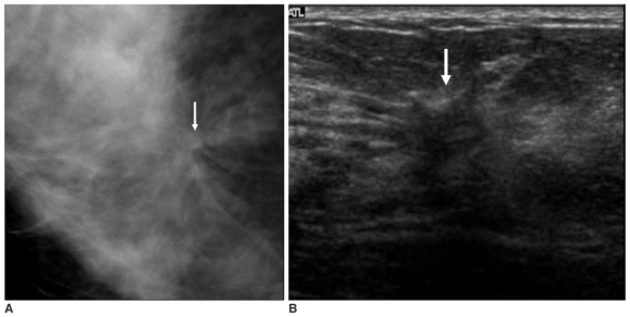
Materials and methods: Sixty-nine women (age range: 25-74 years, mean age: 51.7 years) with 69 papillary lesions (4.9%) were diagnosed and followed after performing sonographically guided 14-gauge core needle biopsies. Surgical excision was performed for 44 (64%) of 69 papillary lesions, and 25 lesions were followed with imaging studies (range: 6-46 months, mean: 17.9 months). The histologic findings upon core biopsy were compared with the surgical, imaging and follow-up findings.
Results: Core needle biopsies of 69 lesions yielded tissue that was classified as benign for 43 lesions, atypical for 18 lesions and malignant for eight lesions. Of the 43 lesions that yielded benign papilloma upon core needle biopsy, one had intraductal papillary carcinoma found upon surgery. An immediate surgical biopsy was recommended for this lesion because of the imaging-histologic discordance. No additional carcinoma was found during the imaging follow-up. Surgical excision was performed for 17 atypical papillary lesions, and this revealed intraductal (n = 6) or invasive (n = 2) papillary carcinoma in 8 (47%) lesions. Of the seven intraductal papillary carcinomas, surgery revealed invasive papillary carcinoma in one (14%).
Conclusion: Our results suggest that papillary lesions of the breast that are diagnosed as benign upon sonographically guided 14-gauge core needle biopsy can be followed when the results are concordant with the imaging findings.
Figures
References
-
- Rosen PP. Benign papillary tumors. In: Rosen PP, editor. Rosen's breast pathology. Philadelphia: Lippincott-Raven; 1997. pp. 67–104.
-
- Rosen PP. Papillary carcinoma. In: Rosen PP, editor. Rosen's breast pathology. Philadelphia: Lippincott-Raven; 1997. pp. 335–354.
-
- Gadd MA. Papillary lesions. In: Harris JR, Lippman ME, Morrow M, Hellman S, editors. Diseases of the breast. Philadelphia: Lippincott-Raven; 1996. pp. 42–45.
-
- Haagensen CD. Solitary intraductal papilloma. In: Haagensen CD, editor. Diseases of the breast. Philadelphia: Saunders; 1986. pp. 136–175.
-
- Ellis IO, Elston CW, Pinder SE. Papillary lesions. In: Elston CW, Ellis IO, editors. Systemic pathology: the breast. 3rd ed. Edinburgh: Churchill Livingstone; 1998. pp. 133–146.
Publication types
MeSH terms
LinkOut - more resources
Full Text Sources
Medical
