

Anterior cruciate ligament tear: reliability of MR imaging to predict stability after conservative treatment
- PMID: 17554192
- PMCID: PMC2627415
- DOI: 10.3348/kjr.2007.8.3.236
Anterior cruciate ligament tear: reliability of MR imaging to predict stability after conservative treatment
Abstract
Objective: The aim of this study is to evaluate the reliability of MR imaging to predict the stability of the torn anterior cruciate ligament (ACL) after complete recovery of the ligament's continuity.
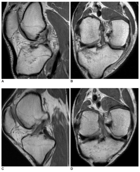
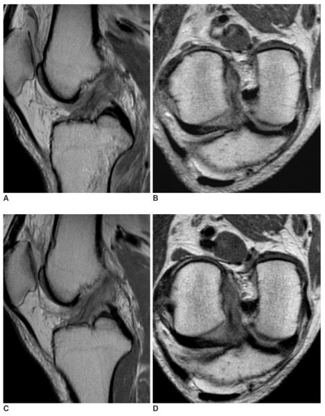
Materials and methods: Twenty patients with 20 knee injuries (13 males and 7 females; age range, 20-54) were enrolled in the study. The inclusion criteria were a positive history of acute trauma, diagnosis of the ACL tear by both the physical examination and the MR imaging at the initial presentation, conservative treatment, complete recovery of the continuity of the ligament on the follow up (FU) MR images and availability of the KT-2000 measurements. Two radiologists, who worked in consensus, graded the MR findings with using a 3-point system for the signal intensity, sharpness, straightness and the thickness of the healed ligament. The insufficiency of ACL was categorized into three groups according to the KT-2000 measurements. The statistic correlations between the grades of the MR findings and the degrees of ACL insufficiency were analyzed using the Cochran-Mantel-Haenszel test (p < 0.05).
Results: The p-values for each category of the MR findings according to the different groups of the KT-2000 measurements were 0.9180 for the MR signal intensity, 1.0000 for sharpness, 0.5038 for straightness and 0.2950 for thickness of the ACL. The MR findings were not significantly different between the different KT-2000 groups.
Conclusion: MR imaging itself is not a reliable examination to predict stability of the ACL rupture outcome, even when the MR images show an intact appearance of the ACL.
Figures
References
-
- Fujimoto E, Sumen Y, Ochi M, Ikuta Y. Spontaneous healing of acute anterior cruciate ligament (ACL) injuries - conservative treatment using an extension block soft brace without anterior stabilization. Arch Orthop Trauma Surg. 2002;122:212–216. - PubMed
-
- Higueras Guerrero V, Torregrosa Andres A, Marti-Bonmati L, Casillas C, Sanfeliu M. Synovialisation of the torn anterior cruciate ligament of the knee: comparison between magnetic resonance and arthroscopy. Eur Radiol. 1999;9:1796–1799. - PubMed
-
- Ihara H, Miwa M, Deya K, Torisu K. MRI of anterior cruciate ligament healing. J Comput Assist Tomogr. 1996;20:317–321. - PubMed
-
- Malanga GA, Giradi J, Nadler SF. The spontaneous healing of a torn anterior cruciate ligament. Clin J Sport Med. 2001;11:118–120. - PubMed
-
- Frank CB. Ligament structure, physiology and function. J Musculoskelet Neuronal Interact. 2004;4:199–201. - PubMed
MeSH terms
LinkOut - more resources
Full Text Sources
Medical
