

Case Reports
doi: 10.3348/kjr.2007.8.3.258.
Primary mucinous adenocarcinoma of a seminal vesicle cyst associated with ectopic ureter and ipsilateral renal agenesis: a case report
Affiliations
- PMID: 17554197
- PMCID: PMC2627409
- DOI: 10.3348/kjr.2007.8.3.258
Item in Clipboard
Case Reports
Primary mucinous adenocarcinoma of a seminal vesicle cyst associated with ectopic ureter and ipsilateral renal agenesis: a case report
Korean J Radiol.
2007 May-Jun.
Abstract
Primary adenocarcinoma of the seminal vesicles is a rare neoplasm. Congenital seminal vesicle cysts are commonly associated with unilateral renal agenesis or dysgenesis. To the best of our knowledge, mucinous adenocarcinoma of the seminal vesicle cyst that's associated with an ectopic ureter opening into the seminal vesicle and ipsilateral renal agenesis has not been described in the radiological literature. We report here on the radiological findings of a primary adenocarcinoma of a seminal vesicle cyst in this condition.
Figures
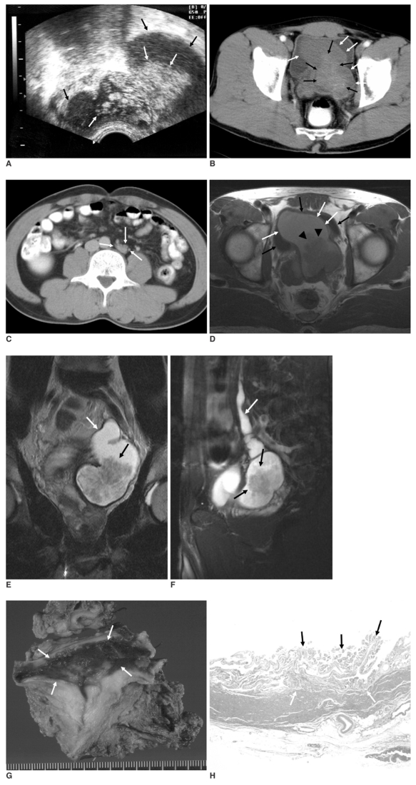
A 41-year-old man with adenocarcinoma of the seminal vesicle cyst associated with an ectopic ureter opening into the seminal vesicle and also ipsilateral renal agenesis. A. Transrectal ultrasonography shows a hyperechoic, intraluminal protruding papillary mass (white arrows) in the cystic change of the left seminal vesicle (black arrows). B. On the contrast-enhanced pelvic CT scan, the papillary solid mass (black arrows) is seen to originate from the wall of the left seminal vesicle cyst (white arrows), and this mass is mildly enhanced. C. A small, abnormal soft tissue density (white arrows) is noted in the aorta's left lateral aspect, suggesting an atrophic or dysgenetic left kidney on the contrast-enhanced abdominal CT at the level of the L4 vertebra. D. An axial T1-weighted MR image shows a large, dilated seminal vesicle (white arrows) with fluid content that has high signal intensity. The cyst contains a low signal intensity papillary mass (arrowheads). The urinary bladder (black arrows) is also noted. E, F. The T2-weighted coronal (E) and sagittal (F) MR images disclose a low signal intensity papillary mass (black arrows) in the left seminal vesicle cyst and a dilated left ectopic ureter (white arrows) draining into the left seminal vesicle. G. The gross specimen shows the internal surface of the seminal vesicle cyst with a residual papillary mass (arrows). The main mass is removed. H. A photomicrograph shows a papillary configuration (black arrows) covered with carcinoma cells without muscular invasion (white arrows) (Hematoxylin & Eosin staining, × 100).
Similar articles
-
[Congenial seminal vesicle cyst with an intracystic papillary adenoma associated with ipsilateral renal agenesis].Radiologe. 2002 Oct;42(10):837-9. doi: 10.1007/s001170100631. Radiologe. 2002. PMID: 12402112 German.
-
Ectopic ureter draining into seminal vesicle cyst: usefulness of MRI.Radiat Med. 1998 Jul-Aug;16(4):309-11. Radiat Med. 1998. PMID: 9814429
-
[Papillary adenocarcinoma in a seminal vesicle cyst associated with contralateral renal agenesis: a case report].Hinyokika Kiyo. 2007 Mar;53(3):175-8. Hinyokika Kiyo. 2007. PMID: 17447487 Japanese.
-
Novel application of da Vinci robotic system in patients of Zinners syndrome--case report and review of literature.Can J Urol. 2010 Apr;17(2):5109-13. Can J Urol. 2010. PMID: 20398450 Review.
-
[Seminal vesicle cyst and ipsilateral kidney failure. Report of 2 cases and review of the literature].Urologe A. 1996 Nov;35(6):490-4. doi: 10.1007/s001200050058. Urologe A. 1996. PMID: 9064889 Review. German.
Cited by
-
Robot-assisted management of Zinner's syndrome: report of seminal vesicle sparing technique and review of literature.J Robot Surg. 2014 Jun;8(2):185-7. doi: 10.1007/s11701-013-0430-3. Epub 2013 Aug 11. J Robot Surg. 2014. PMID: 27637531
-
Primary seminal vesicle adenocarcinoma with a history of seminal vesicle cyst: A case report and review of literature.World J Clin Cases. 2023 May 16;11(14):3261-3266. doi: 10.12998/wjcc.v11.i14.3261. World J Clin Cases. 2023. PMID: 37274041 Free PMC article.
-
Zinner's Syndrome: A Rare Diagnosis of Dysuria Based on Imaging.Case Rep Urol. 2020 Dec 9;2020:8826664. doi: 10.1155/2020/8826664. eCollection 2020. Case Rep Urol. 2020. PMID: 33489407 Free PMC article.
-
Metastatic primary seminal vesicle adenocarcinoma: management of a rare tumour with multiagent chemotherapy and hormonal therapy.BMJ Case Rep. 2017 Oct 10;2017:bcr2017221896. doi: 10.1136/bcr-2017-221896. BMJ Case Rep. 2017. PMID: 29021144 Free PMC article.
-
Zinner syndrome: a radiological journey through a little known condition.Abdom Radiol (NY). 2024 Dec;49(12):4481-4493. doi: 10.1007/s00261-024-04430-5. Epub 2024 Jun 20. Abdom Radiol (NY). 2024. PMID: 38900322 Review.
References
-
- van den Ouden D, Blom JH, Bangma C, de Spiegeleer AH. Diagnosis and management of seminal vesicle cysts associated with ipsilateral renal agenesis: a pooled analysis of 52 cases. Eur Urol. 1998;33:433–440. - PubMed
-
- Schwartz ML, Kenney PJ, Bueschen AJ. Computed tomographic diagnosis of ectopic ureter with seminal vesicle cyst. Urology. 1988;31:55–56. - PubMed
-
- Walsh PC, Retik AB, Vaughan ED, Wein AJ. Campbell's urology. 7th ed. Philadelphia: Saunders; 1998. pp. 3299–3315.
-
- Levisay GL, Holder J, Weigel JW. Ureteral ectopia associated with seminal vesicle cyst and ipsilateral renal agenesis. Radiology. 1975;114:575–576. - PubMed
-
- Matsuki M, Matsuo M, Kaji Y, Okada N. Ectopic ureter draining into seminal vesicle cyst: usefulness of MRI. Radiat Med. 1998;16:309–311. - PubMed
Publication types
MeSH terms
LinkOut - more resources
Full Text Sources
