. 2007 Nov;127(11):2645-55.
doi: 10.1038/sj.jid.5700890. Epub 2007 May 31.

TNF-alpha suppresses alpha-smooth muscle actin expression in human dermal fibroblasts: an implication for abnormal wound healing

Affiliations

TNF-alpha suppresses alpha-smooth muscle actin expression in human dermal fibroblasts: an implication for abnormal wound healing

Mytien T Goldberg et al. J Invest Dermatol. 2007 Nov.

Abstract

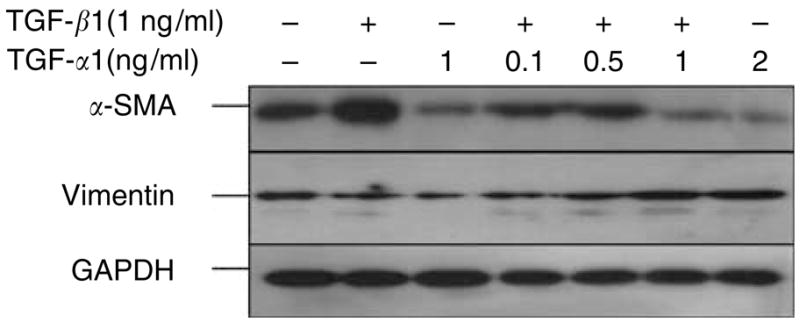
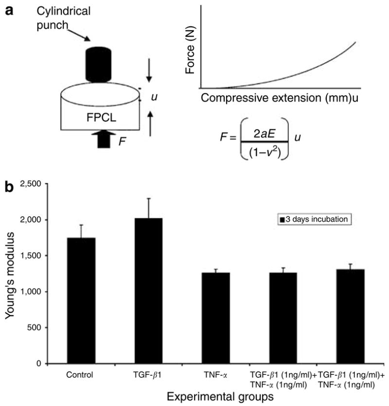
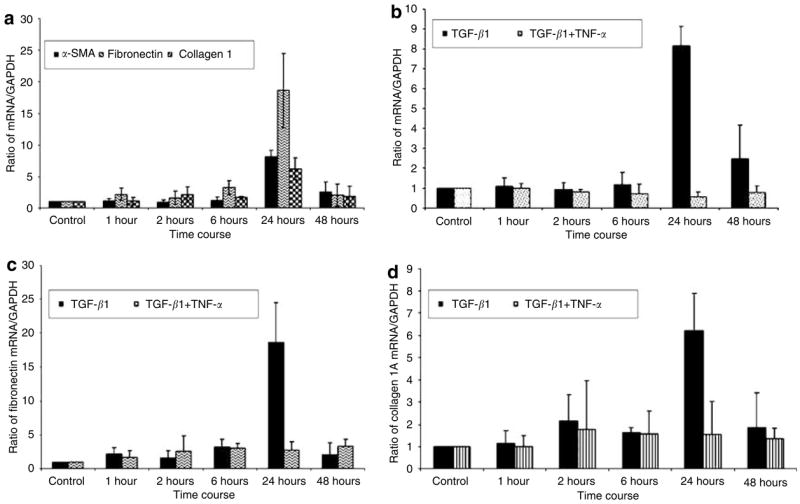
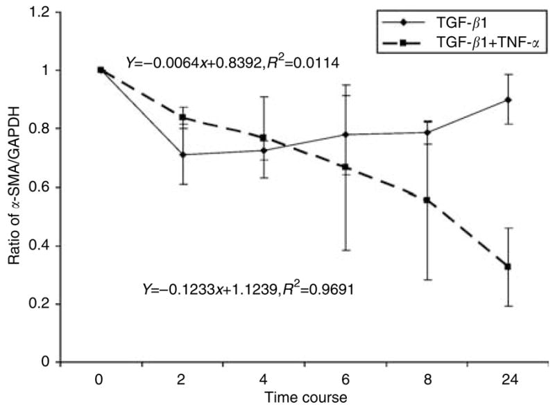
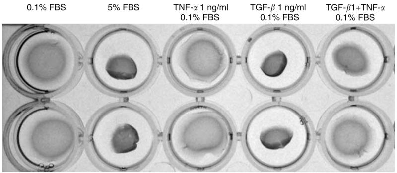
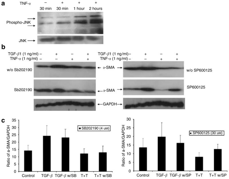
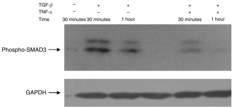
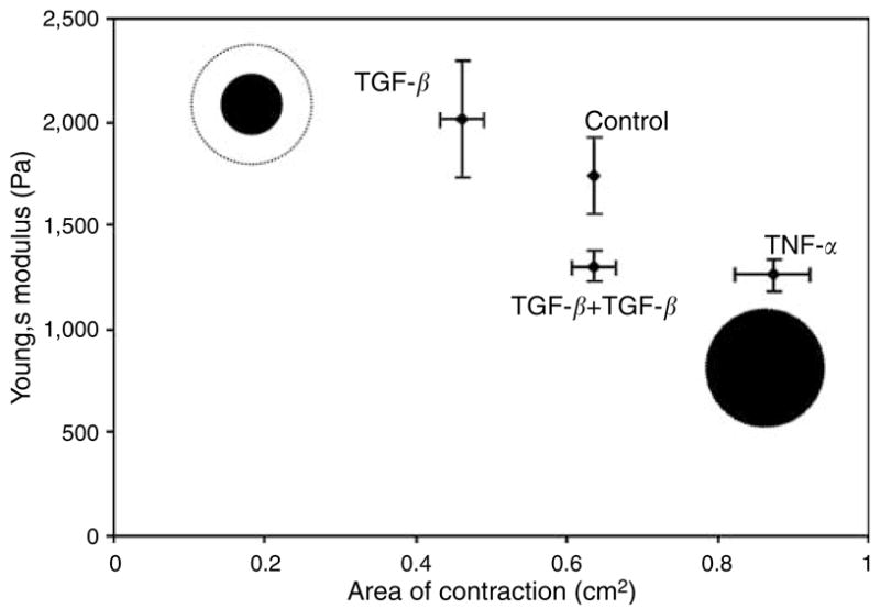
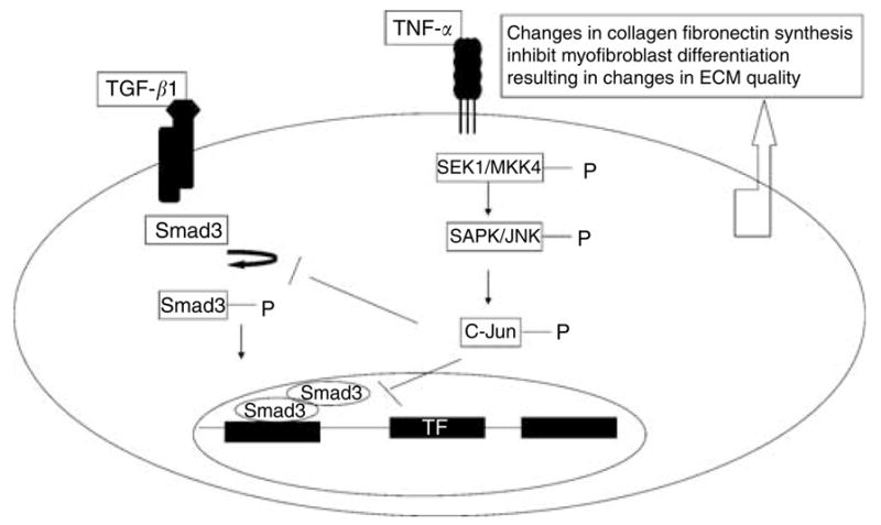
Abnormal wound healing encompasses a wide spectrum, from chronic wounds to hypertrophic scars. Both conditions are associated with an abnormal cytokine profile in the wound bed. In this study, we sought to understand the dynamic relationships between myofibroblast differentiation and mechanical performance of the collagen matrix under tissue growth factor-beta (TGF-beta) and tumor necrosis factor-alpha (TNF-alpha) stimulation. We found TGF-beta increased alpha-smooth muscle actin (alpha-SMA) and TNF-alpha alone decreased the basal alpha-SMA expression. When TGF-beta1 and TNF-alpha were both added, the alpha-SMA expression was suppressed below the baseline. Real-time PCR showed that TNF-alpha suppresses TGF-beta1-induced myofibroblast (fibroproliferative) phenotypic genes, for example, alpha-SMA, collagen type 1A, and fibronectin at the mRNA level. TNF-alpha suppresses TGF-beta1-induced gene expression by affecting its mRNA stability. Our results further showed that TNF-alpha inhibits TGF-beta1-induced Smad-3 phosphorylation via Jun N-terminal kinase signaling. Mechanical testing showed that TNF-alpha decreases the stiffness and contraction of the lattices after 5 days in culture. We proposed that changes in alpha-SMA, collagen, and fibronectin expression result in decreased contraction and stiffness of collagen matrices. Therefore, the balance of cytokines in a wound defines the mechanical properties of the extracellular matrix and optimal wound healing.


Conflict of interest statement

CONFLICT OF INTEREST

The authors state no conflict of interest.

Figures

Figure 1
Figure 1. TNF-α suppresses TGF-β1 induction in human dermal fibroblasts
Normal human dermal fibroblasts (3 × 10⁵ cells/ml) were seeded in collagen I matrix (1 mg/ml). The fibroblast-populated collagen lattices (FPCLs) were treated with TGF-β1 (1 ng/ml), TNF-α (1 ng/ml), or combinations of TGF-β1 (1 ng/ml) and increasing concentrations of TNF-α for 96 hours under 0.1% serum conditions. Representative Western blots for α-SMA, vimentin, and GAPDH are shown.
Figure 2
Figure 2. TNF-α suppresses TGF-β1 promotion of myofibroblast structural elements
(a–d) Representative images of the immunohistochemistry staining for α-SMA. Normal human dermal fibroblasts (1 × 10⁵ cells) were seeded in collagen I matrix (1 mg/ml), which remained attach to the culture plate throughout the cytokine stimulation period. α-SMA was visualized using mouse monoclonal α-SMA FITC conjugated. (a) FPCL under control conditions at 96 hours. (b) FPCL treated with TGF-β1 (1 ng/ml) for 96 hours. (c) FPCL treated with TNF-α (1 ng/ml), and (d) FPCL treated with combination of TGF-β1 (1 ng/ml) and TNF-α (1 ng/ml). Bars = 50 μm (panels a–d). (e–h) Rhodamine-conjugated phalloidin staining for F-actin to examine the assembly of stress fibers. (e) FPCL under control conditions at 96 hours. (f) FPCL treated with TGF-β1 (1 ng/ml) for 96 hours. (g) FPCL treated with TNF-α (1 ng/ml), and (h) FPCL treated with a combination of TGF-β1 (1 ng/ml) plus TNF-α (1 ng/ml). Bars = 50 μm (panels e–h).
Figure 3
Figure 3. TNF-α affects the mechanical response of the fibroblast collagen constructs
The mechanical response of FPCL treated with TGF-β1 (1 ng/ml), TNF-α (1 ng/ml), and a combination of TGF-β1 and TNF-α were measured using compressive indentation assay. In the indentation assay, normal fibroblasts (3 × 10⁵ cells/cm3) were seeded in collagen I matrix (1.5 mg/ml) and induced with cytokines for 72 hours. The constructs underwent indentation measurements. (a) Schematic of the mechanical test and equation used for calculating Young’s modulus. (b) The data are expressed as Young’s modulus for each experimental group.
Figure 4
Figure 4. TNF-α suppresses TGF-β1-induced gene expression at the mRNA level
Fibroblast-populated collagen lattice (FPCL) was prepared at a final cell density of 1.5 × 10⁶ cells/ml in collagen (1 mg/ml) and a 4 ml gel cell mixture was plated in 6 cm2 plate. After polymerization, the FPCL was induced with TGF-β1 (1 ng/ml), TNF-α (1 ng/ml), and TGF-β1 (1 ng/ml) plus TNF-α (1 ng/ml) in DMEM containing 0.1% FBS. Total RNA was extracted and reverse-transcribed into cDNA. cDNA was analyzed for the mRNA expression of α SMA, fibronectin, collagen 1A, and GAPDH by real-time PCR. Results were expressed as a ratio of target gene to GAPDH. Results from three independent experiments are shown with means ± SE (a) Results for TGF-β1 induction of α-SMA, fibronectin, and collagen 1A. (bd) TNF-α suppression of α SMA, fibronectin, and collagen 1A, respectively.
Figure 5
Figure 5. TNF-α affects α-SMA mRNA stability
Adult human dermal fibroblasts were grown in monolayer to confluence and serum-starved for 16 hours before induction with TGF-β1 (1 ng/ml) or TGF-β1 (1 ng/ml) plus TNF-α (1 ng/ml) for 24 hours. At the end of 24 hours, actinomycin D (2.5 μg/ ml) was added and total RNA was harvested at different time points for real-time PCR analysis. The graph represents a ratio of α-SMA and GAPDH mRNA versus time from three independent experiments with SD.
Figure 6
Figure 6. TNF-α abolishes TGF-β1-induced myofibroblast contractility
The stressed fibroblast-populated collagen lattice (FPCL) was prepared in a final cell density of 500,000 cells/ml plus collagen concentration of (1.5 mg/ml) and 0.6 ml volume per gel. The stressed FPCLs were treated under different conditions for 4 days. At the end of treatment, the stressed FPCLs were mechanically released from culture plate and allowed to contract rapidly within 1 hour. The pictures were taken at the end of contraction.
Figure 7
Figure 7. TNF-α decreases TGF-β1 induction of α-SMA expression in a JNK-dependent pathway
(a) Human dermal fibroblasts were grown in monolayer to confluence and serum starved overnight followed by induction with TNF-α. Total protein was harvested at indicated time and Western blot analysis was performed for phosphorylated JNK and JNK proteins. (b) FPCL was prepared in a final cell density of 300,000 cells/ml plus collagen concentration of 1 mg/ml and induced with different cytokine combinations with or without SB202190 and SP600125 inhibitors for 96 hours. Cell lysates were analyzed for α-SMA and GAPDH expression using Western blot. (c) The graph represents mean α-SMA expression as a ratio of GAPDH obtained by densitometric analysis from three independent experiments.
Figure 8
Figure 8. TNF-α inhibits Smad3 phosphorylation in normal human dermal fibroblasts
To test whether TNF-α affects the TGF-β1 Smad signaling pathway, human dermal fibroblasts were grown in monolayer and induced with TGF-β1 (1 ng/ml) or TGF-β1(1 ng/ml) combined with TNF-α (1 ng/ml). Cell lysates were analyzed for phosphorylated Smad3 and GAPDH using Western blot analysis.
Figure 9
Figure 9
Correlation of collagen gel matrix stiffness, seen in indentation assay, with the rate of gel contraction.
Figure 10
Figure 10. A proposed mechanism of TNF-α suppression of TGF-β1-induced gene expression
TNF-α inhibits TGF-β1-induced collagen 1a, fibronectin, and α-SMA expression by inhibiting Smad3 phosphorylation via JNK pathway. Inhibition of Smad3 phosphorylation precluded formation of Smad3 and -4 complex and thus prevented it from regulating the target gene. The overall effect of TNF-α is inhibition of TGF-β1-induced myofibroblast differentiation, leading to softer and less contracted matrix.

References

    1. Abraham DJ, Shiwen X, Black CM, Sa S, Xu Y, Leask A. Tumor necrosis factor alpha suppresses the induction of connective tissue growth factor by transforming growth factor-beta in normal and scleroderma fibro-blasts. J Biol Chem. 2000;275:15220–5. - PubMed
    1. Ashcroft GS, Yang X, Glick AB, Weinstein M, Letterio JL, Mizel DE, et al. Mice lacking Smad3 show accelerated wound healing and an impaired local inflammatory response. Nat Cell Biol. 1999;1:260–6. - PubMed
    1. Bao G, Suresh S. Cell and molecular mechanics of biological materials. Nat Mater. 2003;2:715–25. - PubMed
    1. Connors D, Gies D, Lin H, Gruskin E, Mustoe TA, Tawil NJ. Increase in wound breaking strength in rats in the presence of positively charged dextran beads correlates with an increase in endogenous transforming growth factor-beta1 and its receptor TGF-betaRI in close proximity to the wound. Wound Repair Regen. 2000;8:292–303. - PubMed
    1. Darby I, Skalli O, Gabbiani G. Alpha-smooth muscle actin is transiently expressed by myofibroblasts during experimental wound healing. Lab Invest. 1990;63:21–9. - PubMed

MeSH terms