

Review
doi: 10.1007/s00467-007-0504-4.
Epub 2007 Jun 7.
Molecular regulation of kidney development: is the answer blowing in the Wnt?
Affiliations
- PMID: 17554566
- PMCID: PMC6949197
- DOI: 10.1007/s00467-007-0504-4
Item in Clipboard
Review
Molecular regulation of kidney development: is the answer blowing in the Wnt?
Pediatr Nephrol.
2007 Nov.
Abstract
Development of the metanephric kidney is a complicated process regulated by reciprocal signals from the ureteric bud and the metanephric mesenchyme that regulate tubule formation and epithelial branching morphogenesis. Over the past several years, several studies have suggested that Wnt signaling is involved in multiple aspects of normal kidney development as well as injury response and cancer progression. We will review these data here.
Figures
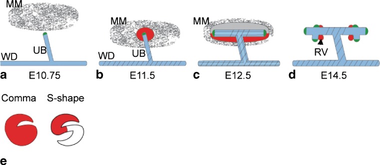
Schematic of kidney development. a At E 10.75, the UB forms from the Wolffian duct (WD). b The UB invades the MM at E 11.5. c The UB forms a T-bud, and the MM condenses. d Branching morphogenesis of the UB takes place, and the renal vesicles (RVs) begin to form. e The RVs will then become comma- and S-shaped bodies, and the branching UB forms the collecting duct. The S-shaped body will fuse to the collecting duct and undergo further morphogenesis to become the nephron (not shown). a–e Expression of Wnts in the developing kidney are indicated as follows: green Wnt11, blue Wnt9b, red Wnt4, diagonal lines Wnt7b
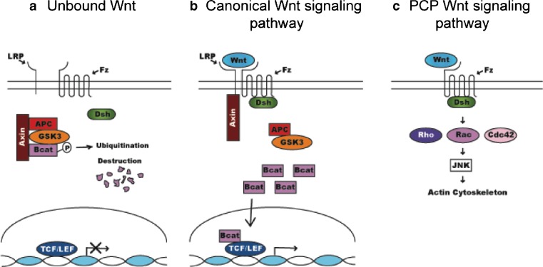
Summary of Wnt signaling. a In the absence of bound Wnt ligand, β-catenin is degraded, due to phosphorylation by GSK-3 beta and binding to the destruction complex. b In canonical signaling, binding of a Wnt to its Fz receptor and Lrp co-receptor results in inactivation of the destruction complex. This allows β-catenin to accumulate in the cytoplasm and translocate into the nucleus, where it activates transcription of Wnt target genes in cooperation with Lef/Tcf co-factors. c In the planar cell polarity (PCP) pathway, Rho, Rac and Cdc42 act downstream of Dsh/Dvl and function to rearrange the actin cytoskeleton and establish cell polarity. In vertebrates, this is thought to be a Wnt-dependent process
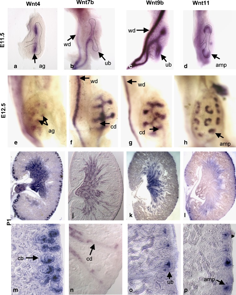
Wnt expression in the developing kidney. The expression pattern of Wnt4 (a, e, i, m), Wnt7b (b, f, j, n), Wnt9b (c, g, k, o), and Wnt11 (d, h, i, p) are shown in the developing kidney by whole-mount in situ hybridization at E 11.5 (a–d) and E 12.5 (e–h). Expression of the Wnts at P1 is shown by section in situ hybridization (i–p). m–p are high-magnification views of the cortex of kidneys shown in i–l. All hybridizations were performed with previously characterized probes and techniques [42]. Wnt7b P1 images provided by Jing Yu. ag aggregate, wd Wolffian duct, ub ureteric bud, amp ureteric bud ampullae, cd collecting duct, cb comma-shaped bodies

Wnt knockout phenotypes. E 14.5 wild-type (WT) (a), Wnt4−/− (b), and Wnt9b−/− (c) kidneys stained with Wnt9b for visualization of the collecting ducts. b In Wnt4−/− kidneys, decreased UB branching occurs. c In Wnt9b−/− kidneys, there is a more severe defect in UB branching than in Wnt4 mutants
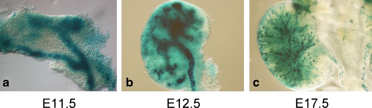
Canonical Wnt signaling in the developing urogenital system. β-galactosidase staining shows canonical Wnt signaling in the developing kidneys of Bat-gal mice. a Staining shows activated β-catenin in the Wolffian duct and UB at E 11.5. At E 12.5 (b) and E 17.5 (c), canonical Wnt signaling is found at high levels in the developing collecting ducts and, perhaps, at lower levels in the MM
References
-
- Vize PD, Woolf AS, Bard JBL, editors. The kidney: from normal development to congenital disease. Boston: Academic Press; 2003.
-
- Duenhoelter JH, Pritchard JA. Fetal respiration. A review. Am J Obstet Gynecol. 1977;129:326–338. - PubMed
-
- Kobayashi A, Kwan KM, Carroll TJ, McMahon AP, Mendelsohn CL, Behringer RR. Distinct and sequential tissue-specific activities of the LIM-class homeobox gene Lim1 for tubular morphogenesis during kidney development. Development. 2005;132:2809–2823. - PubMed
-
- Grobstein C. Some transmission characteristics of the tubule-inducing influence on mouse metanephrogenic mesenchyme. Exp Cell Res. 1957;13:575–587. - PubMed
-
- Grobstein C. Inductive epithlio-mesenchymal interaction in cultured organ rudiments of the mouse metanephros. Science. 1953;118:52–55. - PubMed
Publication types
MeSH terms
Substances
LinkOut - more resources
Full Text Sources
Other Literature Sources
Miscellaneous
