

Optical parameter variability in laser nerve stimulation: a study of pulse duration, repetition rate, and wavelength
- PMID: 17554829
- PMCID: PMC3471085
- DOI: 10.1109/TBME.2007.892925
Optical parameter variability in laser nerve stimulation: a study of pulse duration, repetition rate, and wavelength
Abstract
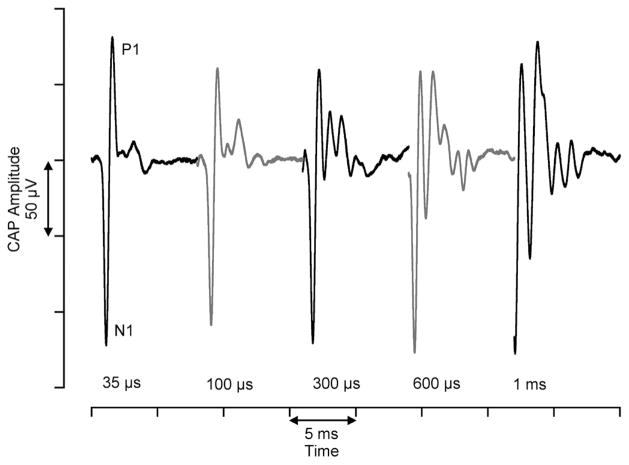
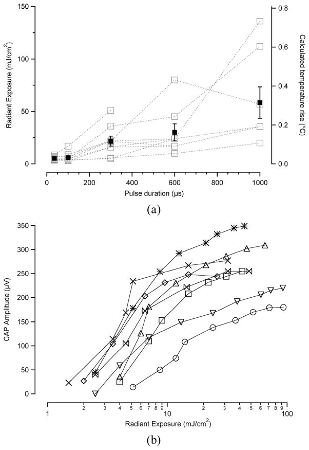
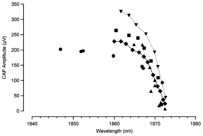
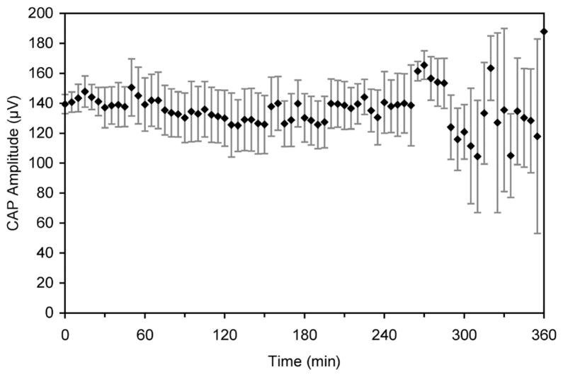
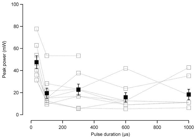
Pulsed lasers can evoke neural activity from motor as well as sensory neurons in vivo. Lasers allow more selective spatial resolution of stimulation than the conventional electrical stimulation. To date, few studies have examined pulsed, mid-infrared laser stimulation of nerves and very little of the available optical parameter space has been studied. In this study, a pulsed diode laser, with wavelength between 1.844-1.873 microm, was used to elicit compound action potentials (CAPs) from the auditory system of the gerbil. We found that pulse durations as short as 35 micros elicit a CAP from the cochlea. In addition, repetition rates up to 13 Hz can continually stimulate cochlear spiral ganglion cells for extended periods of time. Varying the wavelength and, therefore, the optical penetration depth, allowed different populations of neurons to be stimulated. The technology of optical stimulation could significantly improve cochlear implants, which are hampered by a lack of spatial selectivity.
Figures
Similar articles
-
Laser stimulation of auditory neurons: effect of shorter pulse duration and penetration depth.Biophys J. 2008 Apr 15;94(8):3159-66. doi: 10.1529/biophysj.107.117150. Epub 2008 Jan 11. Biophys J. 2008. PMID: 18192375 Free PMC article.
-
Laser stimulation of single auditory nerve fibers.Laryngoscope. 2010 Oct;120(10):2071-82. doi: 10.1002/lary.21102. Laryngoscope. 2010. PMID: 20830761 Free PMC article.
-
Effect of shorter pulse duration in cochlear neural activation with an 810-nm near-infrared laser.Lasers Med Sci. 2017 Feb;32(2):389-396. doi: 10.1007/s10103-016-2129-y. Epub 2016 Dec 20. Lasers Med Sci. 2017. PMID: 27995385
-
Comparison of response properties of the electrically stimulated auditory nerve reported in human listeners and in animal models.Hear Res. 2022 Dec;426:108643. doi: 10.1016/j.heares.2022.108643. Epub 2022 Oct 28. Hear Res. 2022. PMID: 36343534 Free PMC article. Review.
-
Effect of chronic electrical stimulation on cochlear nucleus neuron size in normal hearing kittens.Acta Otolaryngol. 1993 Jul;113(4):489-97. doi: 10.3109/00016489309135851. Acta Otolaryngol. 1993. PMID: 8379304 Review.
Cited by
-
Blind Localization of Heating in Neural Tissues Induced by a Train of the Infrared Pulse Laser.J Lasers Med Sci. 2019 Fall;10(4):264-267. doi: 10.15171/jlms.2019.43. Epub 2019 Oct 1. J Lasers Med Sci. 2019. PMID: 31875117 Free PMC article.
-
Interfaces with the peripheral nervous system for the control of a neuroprosthetic limb: a review.J Neuroeng Rehabil. 2020 Mar 10;17(1):43. doi: 10.1186/s12984-020-00667-5. J Neuroeng Rehabil. 2020. PMID: 32151268 Free PMC article. Review.
-
Attenuated infrared neuron stimulation response in cochlea of deaf animals may associate with the degeneration of spiral ganglion neurons.Biomed Opt Express. 2015 May 7;6(6):1990-2005. doi: 10.1364/BOE.6.001990. eCollection 2015 Jun 1. Biomed Opt Express. 2015. PMID: 26114024 Free PMC article.
-
Identifying optimal parameters for infrared neural stimulation in the peripheral nervous system.Neurophotonics. 2021 Jan;8(1):015012. doi: 10.1117/1.NPh.8.1.015012. Epub 2021 Mar 31. Neurophotonics. 2021. PMID: 33816649 Free PMC article.
-
The cochlear implant: historical aspects and future prospects.Anat Rec (Hoboken). 2012 Nov;295(11):1967-80. doi: 10.1002/ar.22580. Epub 2012 Oct 8. Anat Rec (Hoboken). 2012. PMID: 23044644 Free PMC article. Review.
References
-
- Wells J, Kao C, Mariappan K, Albea J, Jansen ED, Konrad P, Mahadevan-Jansen A. Optical stimulation of neural tissue in vivo. Optics Lett. 2005;31:235–238. - PubMed
-
- Wells J, Kao C, Jansen ED, Konrad P, Mahadevan-Jansen A. Application of infrared light for in vivo neural stimulation. J Biomed Opt. 2005;10(6):064003. - PubMed
-
- Izzo AD, Richter CP, Jansen ED, Walsh JT. Laser stimulation of the auditory nerve. Laser Surg Med. 2006;38(8):745–753. - PubMed
-
- Izzo AD, Suh E, Pathria J, Whitlon DS, Walsh JT, Richter C-P. Selectivity of neural stimulation in the auditory system: A comparison of optic and electric stimuli. J Biomed Opt. 2007;12(2) manuscript #021008. - PubMed
-
- Wells J, Kao C, Konrad P, Mahadevan-Jansen A, Jansen ED. Biophysical mechanisms responsible for pulsed low-level laser excitation of neural tissue. Proc SPIE. 2006;6084
Publication types
MeSH terms
Grants and funding
LinkOut - more resources
Full Text Sources
Other Literature Sources
Medical
Miscellaneous
