. 2007 Jul 3;46(26):7765-80.
doi: 10.1021/bi700425y. Epub 2007 Jun 8.

Human p53 is inhibited by glutathionylation of cysteines present in the proximal DNA-binding domain during oxidative stress

Affiliations

Human p53 is inhibited by glutathionylation of cysteines present in the proximal DNA-binding domain during oxidative stress

Chinavenmeni S Velu et al. Biochemistry. .

Abstract

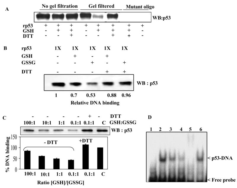
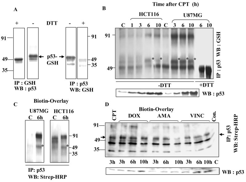
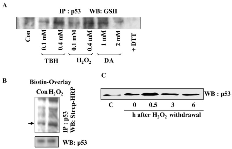
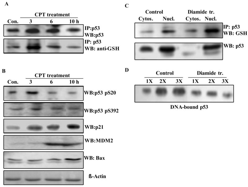
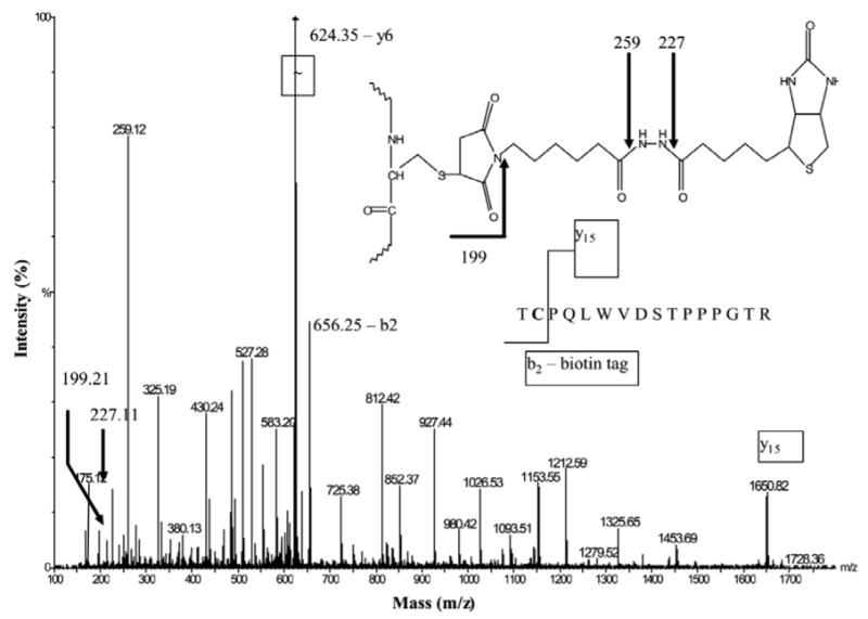
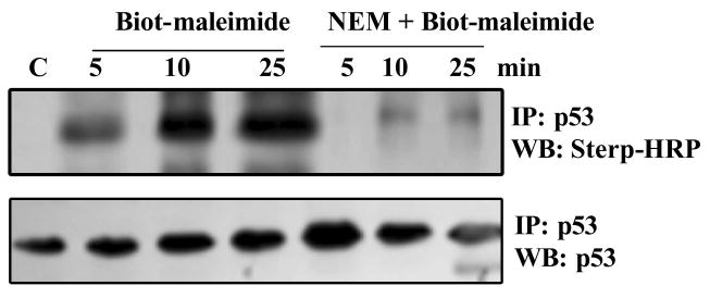
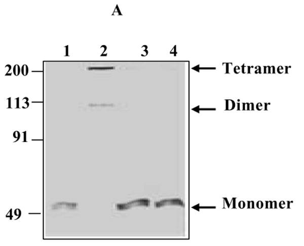
The cellular mechanisms that modulate the redox state of p53 tumor suppressor remain unclear, although its DNA binding function is known to be strongly inhibited by oxidative and nitrosative stresses. We show that human p53 is subjected to a new and reversible posttranslational modification, namely, S-glutathionylation in stressed states, including DNA damage. First, a rapid and direct incorporation of biotinylated GSH or GSSG into the purified recombinant p53 protein was observed. The modified p53 had a significantly weakened ability to bind its consensus DNA sequence. Reciprocal immunoprecipitations and a GST overlay assay showed that p53 in tumor cells was marginally glutathionylated; however, the level of modification increased greatly after oxidant and DNA-damaging treatments. GSH modification coexisted with the serine phophorylations in activated p53, and the thiol-conjugated protein was present in nuclei. When tumor cells treated with camptothecin or cisplatin were subsequently exposed to glutathione-enhancing agents, p53 underwent dethiolation accompanied by detectable increases in the level of p21waf1 expression, relative to the DNA-damaging drugs alone. Mass spectrometry of GSH-modified p53 protein identified cysteines 124, 141, and 182, all present in the proximal DNA-binding domain, as the sites of glutathionylation. Biotinylated maleimide also reacted rapidly with Cys141, implying that this is the most reactive cysteine on the p53 surface. The glutathionylatable cysteines were found to exist in a negatively charged microenvironment in cellular p53. Molecular modeling studies located Cys124 and -141 at the dimer interface of p53 and showed glutathionylation of either residue would inhibit p53-DNA association and also interfere with protein dimerization. These results show for the first time that shielding of reactive cysteines contributes to a negative regulation for human p53 and imply that such an inactivation of the transcription factor may represent an acute defensive response with significant consequences for oncogenesis.


Figures

Figure 1
Figure 1. Human p53 protein is a substrate for glutathionylation in vitro
(A) Rapid incorporation of Biot-GSSG. rp53 (1 μg) was incubated with 5 mM Biot-GSSG in phosphate buffer at 37°C for times shown. Some samples were mixed with 10 mM DTT before loading on non-reducing SDS-Polyacrylamide gels. The blots were reacted with Strep-HRP to obtain the pattern shown. The kinetics of GSSG incorporation remained similar in three independent experiments (P<0.05). Loading of p53 protein on these blots was assessed by western analysis. (B) Kinetics of Biot-GSH incorporation into rp53. rp53 was exposed to 5 mM Biot-GSH, and samples were processed as described in legend to Fig. 1A. (C) p53 glutathionylation is regulated by redox conditions. rp53 protein was treated with mixtures of GSH and GSSG (0.01 to10 mM range) at different ratios in the presence of a constant amount of Biot-GSH (0.4 mM). The samples were incubated at 37°C for 10 min, terminated with the addition of NEM, and processed for blot analysis using Strep-HRP as described above. The lower panel shows the results obtained. DTT reversal of the glutathionation is shown in lane 5. The Blots were reprobed to assess p53 protein loading (upper panel). (D) DAE/NO (NO releasing agent) increases p53 glutathionylation. rp53 was incubated with Biot-GSH in the presence or absence of 0.25 mM DAE/NO, and the labeling was assayed through procedures described above.
Figure 2
Figure 2. GSH or GSSG-modified p53 exhibits significantly decreased binding to consensus DNA sequence
(A) Reduction in the DNA binding activity of rp53 after GSH conjugation. The limited amounts of mercaptoethanol present in rp53 preparations was removed by brief dialysis and used for DNA binding studies described here (See Methods). rp53 was incubated with and without GSH for 30 min. One set of samples (lanes 1-3) were subjected to DNA-affinity immunoblotting assay (DAI) assay without removing the GSH (labeled ‘no gel filtration’). The other set were subjected to gel filtration on Bio-gel P6 spin columns prior to the binding. DAI was performed with biotinylated normal or mutant target sequences for p53. DNA-bound p53 levels are shown. (B) DNA binding assays of rp53 following glutathionylation with GSH or GSSG. rp53 protein at 0.5 μg was incubated with the thiols in sodium phosphate buffer (30 mM, pH 7.5), all samples were then gel-filtered, and DAI assays performed. In some samples, the gel-filtered protein was exposed to DTT before the addition of ds- target DNA. (C) rp53 was incubated with differing GSH/GSSG ratios (100:1 to 0.1:1) for 30 min. All samples were gel-filtered and DAI assays performed. Densitometry of the p53 bands was performed to calculate the % DNA binding shown in the bar graph. Similar results were obtained in three separate experiments (P<0.05). (D) Effect of glutathionylation on the EMSA of p53 binding to its recognition sequence. rp53 was incubated with GSH or GSSG followed by gel-filtration as above, and EMSA performed as described in Methods. Reactions in lanes 1-6 contained 32P-labeled consensus recognition sequence. Lanes: 1, untreated rp53 incubated with mutant oligo; 2, untreated rp53 incubated with consensus oligo; 3, rp53 incubated with GSH; 4, rp53 incubated with GSSG; 5, untreated rp53 incubated with p53 antibody (DO1) before adding the probe to show supershift; 6, rp53 incubated with GSSG, but treated with 5 mM DTT for 10 min prior to probe addition.
Figure 3
Figure 3. Detection of glutathionylated p53 protein in human tumor cells and its upregulation after DNA damage
(A) Evidence from Reciprocal IP /western analysis. HCT116 cells were treated with 25 μM CPT. Extracts (500 μg protein) were immunoprecipitated using antibodies to p53 or GSH. The IPs were electrophoresed under non-reducing conditions, blotted, and reciprocal antibodies were used for p53 detection. Serial dilutions of the cell extracts followed p53 IP provided similar results (not shown). Wherever indicated, the immunoprecipitates were treated with 10 mM DTT for 10 min before SDS-PAGE. DTT-treated sample shows a slightly faster migrating monomer in the left panel. GSH antibodies did not recognize p53 after DTT exposure in the right panel. (B) CPT-induced DNA damage is accompanied by a significant increase in glutathionylated p53 levels. HCT 116 and U87MG cells were treated with 25 μM and 2.5 μM CPT respectively. At each time point, p53 present in 500 μg protein samples was Immunoprecipitated and processed for western analysis using GSH antibodies GSH-linked p53 bands are indicated with an *. C = control. The immunoprecipitates in the last two lanes were treated with DTT before electrophoresis. The lower panel of this figure represents a direct western blot obtained after SDS-PAGE of cell extracts. (C) Detection of GSH-linked p53 by Biot-GST overlay. p53 IPs from control and 6 h CPT-treated U87MG and HCT116 cells were electrophoresed on non-reducing SDS-gels and blotted. The membranes were incubated with Biot-GST protein followed by Strep-HRP staining as described in Methods. (D) Ability of anticancer drugs to generate glutathionylated p53 in U87MG cells. Cells were exposed to doxorubicin (DOX, 10 μM), amsacrine (AMA, 25 μM), vincristine (VINC, 15 μM) for times indicated. p53 present in the cell extracts (500 μg protein) was immunoprecipitated, and the resulting blots were subjected to Biot-GST overlay assay, whose results are shown in the upper panel. The arrows on the left and right point to the position of GSH-linked p53 band, which was not present in the untreated control (Con). The identity of lower band is not known. The lower panel of this figure shows the kinetics of p53 accumulation assessed by western blot analysis of extracts from cells treated with the chemotherapy agents (DOX, AMA, and VINC).
Figure 4
Figure 4. Oxidative stress increases glutathionylated p53 levels in human tumor cells as determined by IP/western, Biot-GST-overlay, and glutathionation-susceptibility assays
(A) Oxidants increase p53 glutathionylation in U87MG cells. Cells were treated with diamide (DA), H2O2 and TBH at concentrations indicated for 15 min. p53 was immunoprecipitated, and the IPs were processed for western analysis using GSH antibodies. In the last lane, p53 IP from 2 mM diamide-treated cells was exposed to 5 mM DTT before electrophoresis. (B) p53 immunoprecipitates from control and H2O2 (0.4 mM, 15 min) treated cells were electrophoresed, and the resulting blot was subjected to Biot-GST overlay assays. Arrow points to the glutathionated p53 band. Direct western blot of p53 is shown in the lower panel. (C) Changes in glutathionylation susceptibility of p53 after H2O2 and DNA-damaging treatments. U87MG cells were exposed to H2O2 (0.4 mM for 15 min) and further postincubated in oxidant-free medium for times shown. Equal protein amounts were then mixed with 0.1 ml GSH-Sepharose beads (ref. 31). All of the bound proteins were eluted with 10 mM DTT, resolved by SDS-PAGE and western blotted for p53.
Figure 5
Figure 5. Glutathionylation coexists with the positive-regulatory phosphorylations in activated p53, the modified protein is nuclear and functionally less active
(A) HCT116 cells were treated with 25 μM CPT up to 10 h. At times specified, p53 present in the cell extracts (300 μg in all samples) was immunoprecipitated. The IPs were electrophoresed on non-reducing SDS-gels, and blotted. Parallel blots were probed using anti-p53 or anti-GSH antibodies. The extracts used for IP were also resolved by SDS-PAGE and blotted. These blots were incubated with p53 phospho-specific antibodies (phosphoserine 20 and phosphoserine 392), or antibodies to hMDM2, p21waf1 and human Bax proteins. The antigens were detected by routine western blotting. (B) Localization of glutathionylated p53 in nuclei. HCT 116 cells were treated with 1 mM diamide for 2 h. The cytosolic and nuclear fractions from control and diamide treated cells were separated using an extraction kit. Equal protein amounts (300 μg) from these samples were subjected to immunoprecipitation of p53 protein. The IPs were electrophoresed and Western blotted using anti-GSH antibodies to obtain the pattern shown in the upper panel. The levels of p53 in the cytosolic and nuclear fractions were assessed by direct Western blotting in the lower panel. (C) Binding of p53 to its consensus recognition sequence in nuclear extracts from diamide-treated cells. HCT116 cells were treated with 1 mM diamide for 3 h. Nuclear extracts were prepared using the Pierce extraction kit. Binding of p53 to its recognition sequence in the presence of increasing nuclear protein, 100 μg (1X), 200 μg (2X) and 300 μg (3X) was assessed by the DNA-affinity immunoblotting (DAI) procedure as described in Experimental Procedures.
Figure 6
Figure 6. Augmentation of intracellular glutathione levels using N-acetylcysteine (NAC) or glutathione ethyl ester (GEE) results in the dethiolation of p53 in cells treated with DNA damaging or oxidizing agents
(A) HCT 116 cells were exposed to 25 μM camptothecin for 2 h to induce p53 glutathionation. These cells and untreated controls were washed, and then incubated with 7.5 mM NAC (found to be optimal in separate experiments) for 5 h. p53 present in equivalent protein amounts (400 μg) was then immunoprecipitated, and western blotted using antibodies to GSH. Cell extracts (50 μg) were immunoblotted in parallel for detection of p21waf1 protein. (B) Following HCT116 cell treatment with tert-butyl hydroperoxide (TBH, 0.2 mM for 15 min) or camptothecin (CPT, 25 μM, 2 h), cells were washed and incubated with 4 mM GEE in the medium for 5 h. p53 immunoprecipitates prepared from cell extracts (400 μg protein) were processed for western blotting for the detection of p53 itself and glutathionated p53. p21waf1 protein levels were assessed by direct western blotting. (C) Following cisplatin treatment (cispt, 20 μM for 2 h), HCT116 cells were incubated in the presence or absence of 7.5 mM NAC for 5 h. Con. = Untreated control cells. P53 immunoprecipitates were western blotted using GSH antibodies for detection of glutathionated p53. Direct immunoblotting was performed for determining p53 and p21waf1 protein levels. (D) Effect of GSH depletion on p53 glutathionation. HCT116 cells were treated with buthionine sulfomine (BSO, 100 μM for 20 h). p53 immunoprecipitates from control and BSO-treated cells were western blotted using antibodies to GSH.
Figure 7
Figure 7. QTOF MS/MS spectrum of T8 biotinylated peptide
rp53 was treated with biotin-maleimide, trypsinized, and the biotin containing peptides were purified on monomeric avidin resin (32). The doubly protonated molecular ion at m/z = 1153.03 (T8 peptide of p53, 140TCPVQLWVDSTPPPGTR156) was fragmented in the collision cell of a QTOF instrument under low-energy conditions (42 eV). Three fragmentation ions (m/z = 199.2, 227.1 and 259.1) indicate the presence of the biotin tag pictured in the inset. The presence of the y15 ion m/z = (1649.80) and a b2 (m/z = 656.25) ion containing the whole biotin-maleimide tag, locates the biotin tag on Cys141.
Figure 8
Figure 8. Rapid incorporation of Biot-maleimide into cellular p53
Biotin-labeled maleimide incorporation into the endogenous p53 protein in HCT116 cells was determined as described in Methods. Briefly, HCT116 cell extracts were exposed to 5mM Biot-maleimide with or without NEM pretreatment. 5 mM NEM was also added to terminate the reactions. p53 immunoprecipitates were prepared from these extracts and western blotted using the Strep-HRP reagent.
Figure 9
Figure 9. Protein cross-linking and structural modeling of S- glutathionated p53
(A) Glutaraldehyde cross-linking and gel analysis of the oligomeric structure of glutathionated p53. rp53 (5 μg) was treated with 5 mM GSH or GSSG for 1 h. The thiols present in the samples were removed by gel-filtration on Bio-gel P6 spin columns. Glutaraldehyde was then added to the eluted proteins to 0.1% concentration, and incubated for 10 min. Next, SDS-sample buffer containing 2-mercaptoethanol was added, and the samples were resolved by SDS-PAGE, and the gel stained with Comassie Blue. Lanes: 1, untreated rp53; 2, cross-linked rp53; 3, rp53 incubated with GSH, and then cross-linked; 4, rp53 incubated with GSSG, and then cross-linked. (B) A ribbon representation of the crystal structure of p53-DNA complex. The p53 chains A and B found in the crystal structure (1TSR) are colored magenta and yellow respectively. These monomers form a dimer and stably associate with the consensus sequence in the crystal structure. Cys124 and Cys141 are present at the dimerization interface and are shown as color coded ball- and –sticks. Note the close proximity of these residues in the native structure. (C) Schematic ribbon representation of the energy-minimized model of p53 glutathionated at Cys124. Procedures used for protein modeling are described in Experimental Procedures. Structural alterations due to the modification resulted in the retraction of the DNA presenting loop that harbors Lys120 in the B monomer (yellow, displaced loop marked with **). Changes also occurred at the dimer interface loop, and elsewhere in the molecule (some are marked with an *). The deformation at the protein interface was determined to inhibit the dimerization. Modeling of a glutathione to Cys141 induced similar perturbations, and the spatial closeness of cysteines 124 and 141 indicated that both may not be thiolated in a single molecule.

References

    1. Mills AA. p53: link to the past bridge to the future. Genes Develop. 2005;19:2091–2099. - PubMed
    1. Inoue T, Wu L, Stuart J, Maki CG. Control of p53 nuclear accumulation in stressed cells. FEBS Lett. 2005;579:4978–4984. - PubMed
    1. Strosznajder RP, Jesko H, Banasik M, Tanaka S. Effects of p53 inhibitor on survival and death of cells subjected to oxidative stress. J Physiol Pharmacol. 2005;4:215–221. - PubMed
    1. Bode AM, Dong Z. Post-translational modification of p53 in tumorigenesis. Nat Rev Cancer. 2004;4:793–805. - PubMed
    1. Martindale JL, Holbrook NJ. Cellular response to oxidative stress: signaling for suicide and survival. J Cellular Physiol. 2002;192:1–15. - PubMed

Publication types

MeSH terms