. 2007 Sep 1;406(2):285-95.
doi: 10.1042/BJ20070479.

Mutant alpha-galactosidase A enzymes identified in Fabry disease patients with residual enzyme activity: biochemical characterization and restoration of normal intracellular processing by 1-deoxygalactonojirimycin

Affiliations

Mutant alpha-galactosidase A enzymes identified in Fabry disease patients with residual enzyme activity: biochemical characterization and restoration of normal intracellular processing by 1-deoxygalactonojirimycin

Satoshi Ishii et al. Biochem J. .

Abstract

Fabry disease is a lysosomal storage disorder caused by the deficiency of alpha-Gal A (alpha-galactosidase A) activity. In order to understand the molecular mechanism underlying alpha-Gal A deficiency in Fabry disease patients with residual enzyme activity, enzymes with different missense mutations were purified from transfected COS-7 cells and the biochemical properties were characterized. The mutant enzymes detected in variant patients (A20P, E66Q, M72V, I91T, R112H, F113L, N215S, Q279E, M296I, M296V and R301Q), and those found mostly in mild classic patients (A97V, A156V, L166V and R356W) appeared to have normal K(m) and V(max) values. The degradation of all mutants (except E59K) was partially inhibited by treatment with kifunensine, a selective inhibitor of ER (endoplasmic reticulum) alpha-mannosidase I. Metabolic labelling and subcellular fractionation studies in COS-7 cells expressing the L166V and R301Q alpha-Gal A mutants indicated that the mutant protein was retained in the ER and degraded without processing. Addition of DGJ (1-deoxygalactonojirimycin) to the culture medium of COS-7 cells transfected with a large set of missense mutant alpha-Gal A cDNAs effectively increased both enzyme activity and protein yield. DGJ was capable of normalizing intracellular processing of mutant alpha-Gal A found in both classic (L166V) and variant (R301Q) Fabry disease patients. In addition, the residual enzyme activity in fibroblasts or lymphoblasts from both classic and variant hemizygous Fabry disease patients carrying a variety of missense mutations could be substantially increased by cultivation of the cells with DGJ. These results indicate that a large proportion of mutant enzymes in patients with residual enzyme activity are kinetically active. Excessive degradation in the ER could be responsible for the deficiency of enzyme activity in vivo, and the DGJ approach may be broadly applicable to Fabry disease patients with missense mutations.


Figures

Figure 1
Figure 1. pH Stability of wild-type and mutant α-Gal A enzymes
The enzymes were purified from cell lysates of COS-7 cells transfected with expression plasmids encoding various mutant α-Gal A enzymes. The stable pH range is defined as the pH in which more than 50% enzyme activity remained after incubation at 37 °C for 1 h in the absence (dark grey bar) or presence (light grey bar) of 1 μM DGJ.
Figure 2
Figure 2. Effects of ERAD inhibitors on the amount of mutant α-Gal A expressed in COS-7 cells
Wild-type or mutant α-Gal A enzymes were transiently expressed with FuGENE™ 6 transfection reagent in COS-7 cells. At 5 h after transfection, cells were treated with 2 μM lactacystin (LC), 0.2 mM kifunensine (KFN) or nothing as a control (C), and harvested at 48 h. Western blot analyses of cell lysates from transfected COS-7 cells were performed.
Figure 3
Figure 3. Effect of DGJ treatment on various mutant α-Gal A enzymes expressed in COS-7 cells
COS-7 cells were re-plated on six-well plates and transfected with expression constructs containing various mutant α-Gal A cDNAs using Lipofectamine™ 2000 reagent. Cells were cultured in complete medium for 3 days with or without 20 μM DGJ. α-Gal A activity and Western blot analyses of cell lysates from transfected COS-7 cells were performed as described in the Materials and methods section. Lane 1, Mock transfection; lane 2, wild-type α-Gal A; lane 3, A20P; lane 4, E59K; lane 5, E66Q; lane 6, M72V; lane 7, I91T; lane 8, A97V; lane 9, R112H; lane 10, F113L; lane 11, P146S; lane 12, A156V; lane 13, L166V; lane 14, N215S; lane 15, Q279E; lane 16, M296I; lane 17, M296V; lane 18, R301Q; lane 19, R356W; lane 20, G373D; and lane 21, G373S.
Figure 4
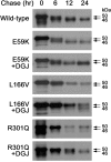
Figure 4. Metabolic labelling of mutant α-Gal A expressed in COS-7 cells
COS-7 cells were transfected in six-well plates with expression plasmids coding for the wild-type, E59K, L166V or R301Q α-Gal A enzymes respectively, and cultured in the absence or presence of 20 μM DGJ. The cells were exposed to 4 μl of [35S]Protein labelling mix for 2 h. After washing the cells with PBS, the labelled proteins were chased by replacement of medium for the indicated period. Following immunoprecipitation with a polyclonal anti-α-Gal A antibody, an aliquot (one-fifth of the sample) was analysed by SDS/PAGE (10% gels) and visualized by fluorography.
Figure 5
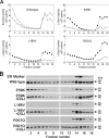
Figure 5. Subcellular fractionation of the wild-type or mutant α-Gal A expressed in COS-7 cells
COS-7 cells were transfected with expression plasmids pCXN2-GLA, pCXN2-GLA-E59K, pCXN2-GLA-L166V or pCNX2-GLA-R301Q respectively. Subcellular fractionation was performed by Percoll gradient centrifugation at 25000 g for 1 h. Each fraction was collected, and the density determined using density markers (Fraction 2, 1.11 g/ml; fraction 4, 1.07 g/ml; fraction 7, 1.056 g/ml; fraction 12, 1.053 g/ml; and fraction 16, 1.041 g/ml). (A) α-Gal A (○) and β-hexosaminidase (∇) activities in each fraction of wild type α-Gal A. α-Gal A activities of E59K, L166V and R301Q in cells treated with 20 μM DGJ (●) or without (○) in each fraction. (B) Western blot analyses with an antibody against human α-Gal A. Bip was used as an ER marker protein.
Figure 6
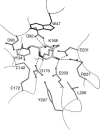
Figure 6. Predicted interactions between DGJ and the active site of α-Gal A
Based upon the crystal structure of α-Gal A with bound α-galactose, we modelled the interactions of α-Gal A with DGJ. The DGJ is shown bound to the active-site of the enzyme, in a manner very similar to α-galactose binding. The key interactions to the 2, 3, 4, and 6 hydroxy groups on the ligand are maintained when either α-galactose or DGJ binds to the active-site. One key interaction between Glu231 on the enzyme and the 1 hydroxy group of α-galactose is lost when DGJ binds, because DGJ lacks a functional group at the 1 position. At acidic pH, DGJ becomes protonated and positively charged, which may increase its affinity for the highly negatively charged enzyme.

References

    1. Brady O. R., Gal A. E., Bradley R. M., Martensson E., Warshaw A. L., Laster L. Enzymatic defect in Fabry's disease: ceramidetrihexosidase deficiency. N. Engl. J. Med. 1967;276:1163–1167. - PubMed
    1. Desnick R. J., Ioannou Y. A., Eng C. M. The Metabolic and Molecular Bases of Inherited Disease. In: Scriver C. R., Beaudet A. L., Sly W. S., Valle D., editors. New York: McGraw-Hill; 2001. pp. 3733–3774.
    1. Shah J. S., Elliott P. M. Fabry disease and the heart: an overview of the natural history and the effect of enzyme replacement therapy. Acta Paediatr. Suppl. 2005;94:11–14. discussion 9–10. - PubMed
    1. Branton M., Schiffmann R., Kopp J. B. Natural history and treatment of renal involvement in Fabry disease. J. Am. Soc. Nephrol. 2002;13(Suppl. 2):S139–S143. - PubMed
    1. Bishop D. F., Grabowski G. A., Desnick R. J. Fabry disease: an asymptomatic hemizygote with significant residual α-galactosidase A activity. Am. J. Hum. Genet. 1981;33:71A.

Publication types

MeSH terms