. 2008 Dec;29(12):1795-804.
doi: 10.1016/j.neurobiolaging.2007.04.023. Epub 2007 Jun 6.

Regulation of the NMDA receptor-mediated synaptic response by acetylcholinesterase inhibitors and its impairment in an animal model of Alzheimer's disease

Affiliations

Regulation of the NMDA receptor-mediated synaptic response by acetylcholinesterase inhibitors and its impairment in an animal model of Alzheimer's disease

Guojun Chen et al. Neurobiol Aging. 2008 Dec.

Abstract

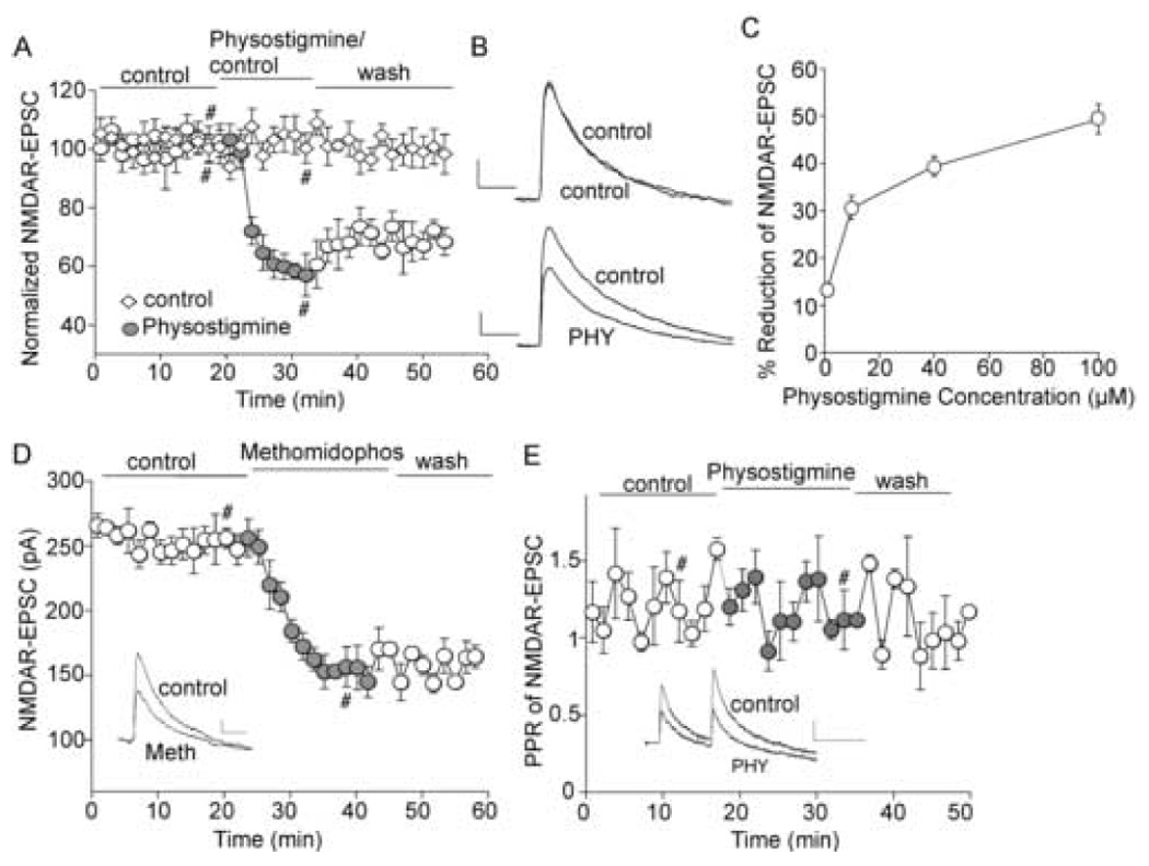
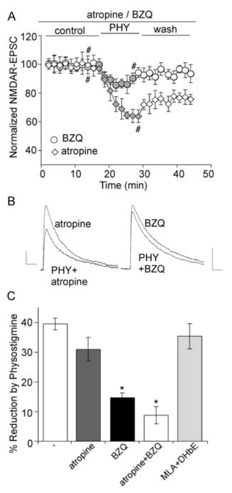
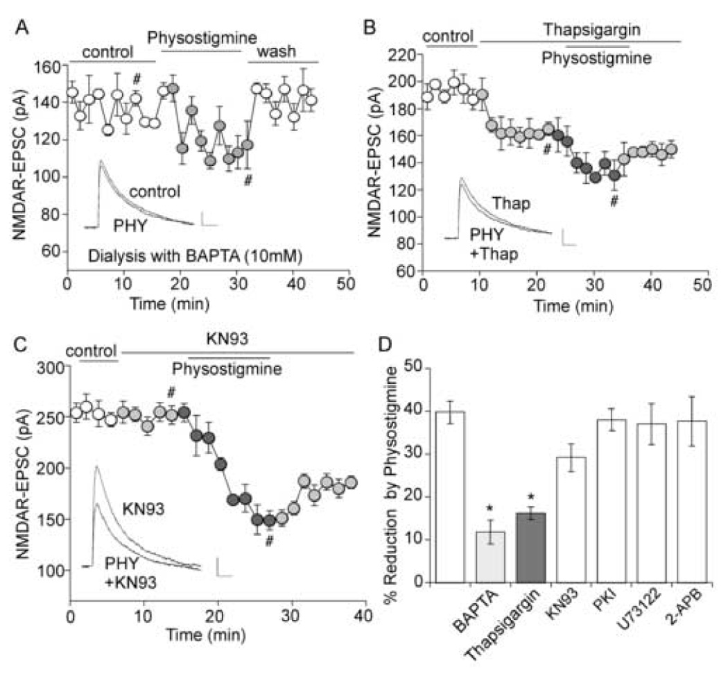
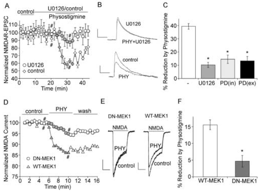
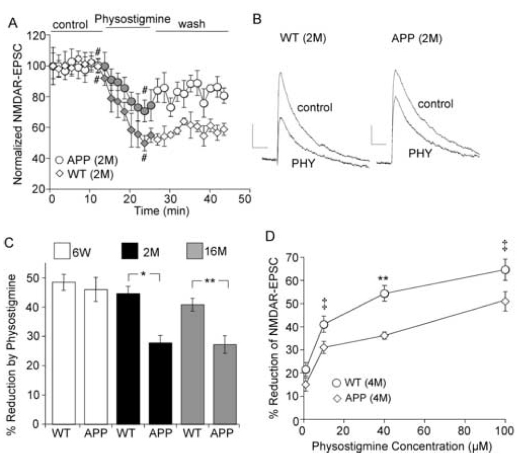
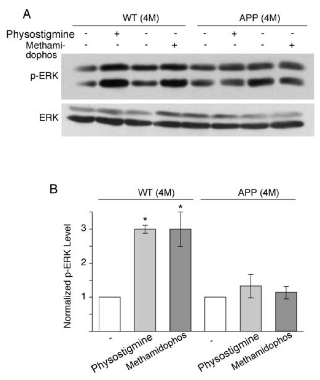
The cholinergic system is crucial for cognitive processes and the deficient acetylcholine (ACh) function has been implicated in Alzheimer's disease (AD). Inhibitors of acetylcholinesterase (AChE), which act to enhance cholinergic function by prolonging the action of endogenously released ACh, have been used as the major therapy of AD. To understand the functional roles of cholinergic enhancement in prefrontal cortex (PFC), a key brain region for cognition, we examined the impact of AChE inhibitors in PFC neurons on synaptic responses mediated by the NMDA receptor (NMDAR), an important player in learning and memory. We found that AChE inhibitors produced a strong and persistent reduction of the amplitude of NMDA receptor-mediated excitatory postsynaptic current (NMDAR-EPSC). This effect was mainly mediated by nicotinic ACh receptors, and through a Ca(2+)-dependent mechanism. Inhibition of extracellular signal-regulated kinases (ERK) abolished the regulation of NMDAR function by AChE inhibitors, suggesting the involvement of ERK. In the transgenic mouse model of AD overexpressing mutant beta-amyloid precursor protein (APP), the effect of AChE inhibitors on NMDAR-EPSC was significantly impaired, which was associated with their diminished effect on ERK activation. Taken together, these results suggest that one of the key targets of endogenous ACh involved in cognition is the NMDAR-mediated transmission. Loss of the regulation of synaptic NMDAR responses by endogenous ACh may contribute to the cognitive deficiency in AD.


Conflict of interest statement

Disclosure Statement

All authors have no actual or potential conflicts of interest with other people or organizations within three years of beginning the work presented here.

Figures

Figure 1
Figure 1. AChE inhibitors cause a reduction of NMDAR-EPSC amplitudes in cortical pyramidal neurons
A, Plot of normalized peak NMDAR-EPSC amplitudes as a function of time and drug (the AChE inhibitor physostigmine, 40 µM, or control ACSF) application in two representative PFC pyramidal neurons. B. Representative traces of NMDAR-EPSC from the records used to construct A (at time points denoted by #). Scale bars: 20pA/100ms. C. Dose response curve showing the effect of physostigmine on NMDAR-EPSC. D. Plot of peak NMDAR-EPSC amplitudes as a function of time and methomidophos (50 µM, another AChE inhibitor) application in a representative PFC pyramidal neuron. E. Plot of paired pulse ratio (PPR) of NMDAR-EPSC as a function of time and drug application. Inset (D, E), representative traces of NMDAR-EPSC (at time points denoted by #). Scale bars: 20pA/100ms.
Figure 2
Figure 2. The reduction of NMDAR-EPSC by physostigmine is mainly mediated by nicotinic acetylcholine receptors
A. Plot of normalized NMDAR-EPSC amplitudes as a function of time and physostigmine (40 µM) application in neurons treated with either atropine (50 µM) or benzoquinonium (BZQ, 50 µM). B. Current traces taken from the records used to construct A (at time points denoted by #). Scale bars: 10pA/100ms. C. Bar plot summary showing the percentage reduction of NMDAR-EPSC by physostigmine in the presence of atropine, BZQ, atropine + BZQ, or MLA (10 µM) + DHβE (0.1 µM). *: p < 0.001, t-test.
Figure 3
Figure 3. The effect of physostigmine on NMDAR-EPSC is through a mechanism depending on Ca2+
A. Plot of NMDAR-EPSC as a function of time and physostigmine (40 µM) application in a neuron loaded with BAPTA (10 mM). Inset, representative current traces (at time points denoted by #). Scale bars: 20pA/100ms. B, C. Plot of NMDAR-EPSC as a function of time and drug application. Note that thapsigargin (10 µM, B), but not KN93 (10 µM, C), prevented the effect of physostigmine. Inset (B, C), representative current traces (at time points denoted by #). Scale bars: 10pA/100ms. D. Bar plot summary showing the percentage reduction of NMDAR-EPSC by physostigmine in neurons treated with different agents. *: p < 0.005, t-test.
Figure 4
Figure 4. ERK is involved in the regulation of NMDAR current by physostigmine
A. Plot of normalized NMDAR-EPSC as a function of time and physostigmine (40 µM) application in the absence (control) or presence of U0126 (20 µM). B. Representative current traces taken from records used to construct A (at time points denoted by #). Scale bars: 20pA/100ms. C. Bar plot summary of the percentage reduction of NMDAR-EPSC by physostigmine in neurons treated without or with U0126 or PD98059 (25 µM internal or external application). *: p < 0.001, t-test. D. Plot of normalized NMDA (100 µM)-evoked currents as a function of time and physostigmine (40 µM) application in cultured cortical neurons transfected with either wild type MEK1 (WT-MEK1) or dominant negative MEK1 (DN-MEK1). E. Representative current traces taken from records used to construct D (at time points denoted by #). Scale bars: 50pA/0.5sec. F. Bar plot summary of the percentage reduction of NMDAR current by physostigmine in neurons transfected with WT-MEK1 or DN-MEK1. *: p < 0.001, t-test.
Figure 5
Figure 5. The regulation of NMDAR-EPSC by physostigmine is impaired in APP transgenic mice
A. Plot of normalized NMDAR-EPSC as a function of time and physostigmine (40 µM) application in cortical pyramidal neurons from a 2-month-old wild type mouse (WT) and a 2-month-old APP transgenic mouse (APP). B. Representative current traces taken from records used to construct A (at time points denoted by #). Scale bars: 20pA/100ms. C. Bar plot summary of the percentage reduction of NMDAR-EPSC by physostigmine in wild-type and APP transgenic mice at different ages (6 weeks, 2 months, and 16 months). *: p < 0.001; **: p < 0.005, t-test. D. Dose response curves showing the effect of physostigmine on NMDAR-EPSC in 4-month-old WT and APP transgenic mice. ‡: p < 0.01; **: p < 0.005, t-test.
Figure 6
Figure 6. The ERK activation by AChE inhibitors is diminished in APP transgenic mice
A. Representative Western blots of phospho-ERK and total ERK in cortical slices treated without or with AChE inhibitors (physostigmine: 40 µM; methomidophos: 40 µM) from WT and APP transgenic mice (4-month-old). B. Bar plot summary showing the phospho-ERK levels in cortical slices from WT and APP mice under different treatments. *: p < 0.005, t-test.

References

    1. Akasofu S, Kimura M, Kosasa T, Ogura H, Sawada K. Protective effect of donepezil in primary-cultured rat cortical neurons exposed to N-methyl-d-aspartate (NMDA) toxicity. Eur J Pharmacol. 2006;530(3):215–222. - PubMed
    1. Auld DS, Kar S, Quirion R. Beta-amyloid peptides as direct cholinergic neuromodulators: a missing link? Trends Neurosci. 1998;21(1):43–49. - PubMed
    1. Bartus RT, Dean RL, 3rd, Beer B, Lippa AS. The cholinergic hypothesis of geriatric memory dysfunction. Science. 1982;217(4558):408–414. - PubMed
    1. Benzi G, Moretti A. Is there a rationale for the use of acetylcholinesterase inhibitors in the therapy of Alzheimer's disease? Eur J Pharmacol. 1998;346(1):1–13. - PubMed
    1. Blusztajn JK, Berse B. The cholinergic neuronal phenotype in Alzheimer's disease. Metab Brain Dis. 2000;15(1):45–64. - PubMed

Publication types

MeSH terms

Substances