

Nanoparticle effects on rat alveolar epithelial cell monolayer barrier properties
- PMID: 17555923
- PMCID: PMC3855017
- DOI: 10.1016/j.tiv.2007.04.003
Nanoparticle effects on rat alveolar epithelial cell monolayer barrier properties
Abstract
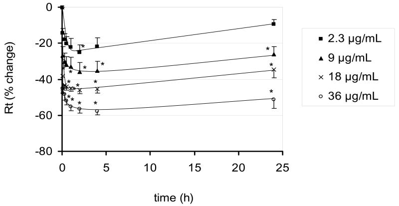
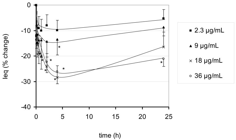
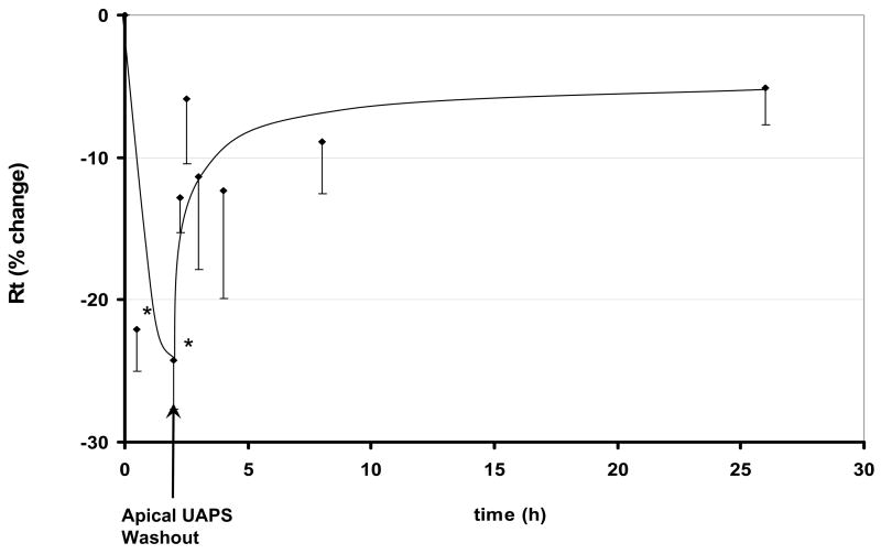
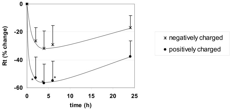
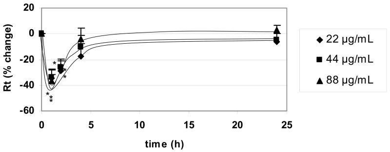
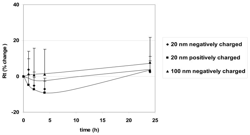
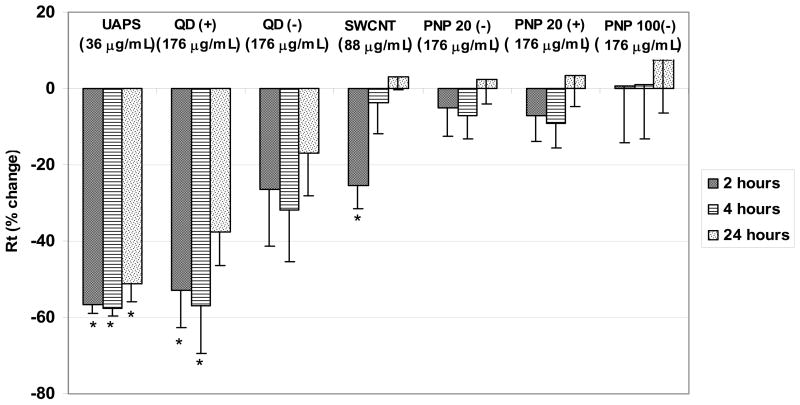
Inhaled nanoparticles have been reported to contribute to deleterious effects on human health. In this study, we investigated the effects of ultrafine ambient particulate suspensions (UAPS), polystyrene nanoparticles (PNP; positively and negatively charged; 20, 100, 120 nm), quantum dots (QD; positively and negatively charged; 30 nm) and single-wall carbon nanotubes (SWCNT) on alveolar epithelial cell barrier properties. Transmonolayer resistance (R(t)) and equivalent short-circuit current (I(eq)) of primary rat alveolar epithelial monolayers were measured in the presence and absence of varying concentrations of apical nanoparticles. In some experiments, apical-to-basolateral fluxes of radiolabeled mannitol or inulin were determined with or without apical UAPS exposure and lactate dehydrogenase (LDH) release was analyzed after UAPS or SWCNT exposure. Results revealed that exposure to UAPS decreased R(t) and I(eq) significantly over 24 h, although neither mannitol nor inulin fluxes changed. Positively charged QD decreased R(t) significantly (with subsequent recovery), while negatively charged QD did not. R(t) decreased significantly after SWCNT exposure (with subsequent recovery). On the other hand, PNP exposure had no effects on R(t) or I(eq). No significant increases in LDH release were observed after UAPS or SWCNT exposure. These data indicate that disruption of alveolar epithelial barrier properties due to apical nanoparticle exposure likely involves alteration of cellular transport pathways and is dependent on specific nanoparticle composition, shape and/or surface charge.
Figures
Similar articles
-
Translocation of PEGylated quantum dots across rat alveolar epithelial cell monolayers.Int J Nanomedicine. 2011;6:2849-57. doi: 10.2147/IJN.S26051. Epub 2011 Nov 10. Int J Nanomedicine. 2011. PMID: 22131830 Free PMC article.
-
Alveolar epithelial cell injury due to zinc oxide nanoparticle exposure.Am J Respir Crit Care Med. 2010 Dec 1;182(11):1398-409. doi: 10.1164/rccm.201002-0185OC. Epub 2010 Jul 16. Am J Respir Crit Care Med. 2010. PMID: 20639441 Free PMC article.
-
Polystyrene nanoparticle trafficking across alveolar epithelium.Nanomedicine. 2008 Jun;4(2):139-45. doi: 10.1016/j.nano.2008.02.002. Epub 2008 Mar 28. Nanomedicine. 2008. PMID: 18375191
-
Nanoparticle translocation across mouse alveolar epithelial cell monolayers: species-specific mechanisms.Nanomedicine. 2013 Aug;9(6):786-94. doi: 10.1016/j.nano.2013.01.007. Epub 2013 Feb 20. Nanomedicine. 2013. PMID: 23454523 Free PMC article.
-
Significance of particle parameters in the evaluation of exposure-dose-response relationships of inhaled particles.Inhal Toxicol. 1996;8 Suppl:73-89. Inhal Toxicol. 1996. PMID: 11542496 Review.
Cited by
-
Tight junction between endothelial cells: the interaction between nanoparticles and blood vessels.Beilstein J Nanotechnol. 2016 May 6;7:675-84. doi: 10.3762/bjnano.7.60. eCollection 2016. Beilstein J Nanotechnol. 2016. PMID: 27335757 Free PMC article. Review.
-
Pulmonary applications and toxicity of engineered nanoparticles.Am J Physiol Lung Cell Mol Physiol. 2008 Sep;295(3):L400-11. doi: 10.1152/ajplung.00041.2008. Epub 2008 Jul 18. Am J Physiol Lung Cell Mol Physiol. 2008. PMID: 18641236 Free PMC article. Review.
-
Airborne micro- and nanoplastics: emerging causes of respiratory diseases.Part Fibre Toxicol. 2024 Dec 4;21(1):50. doi: 10.1186/s12989-024-00613-6. Part Fibre Toxicol. 2024. PMID: 39633457 Free PMC article. Review.
-
In vitro effects of silver nanoparticles on the mitochondrial respiratory chain.Mol Cell Biochem. 2010 Sep;342(1-2):51-6. doi: 10.1007/s11010-010-0467-9. Epub 2010 Apr 22. Mol Cell Biochem. 2010. PMID: 20411305
-
Mechanisms of alveolar epithelial translocation of a defined population of nanoparticles.Am J Respir Cell Mol Biol. 2010 May;42(5):604-14. doi: 10.1165/rcmb.2009-0138OC. Epub 2009 Jul 2. Am J Respir Cell Mol Biol. 2010. PMID: 19574531 Free PMC article.
References
-
- Adamson IY, Bowden DH. Derivation of type 1 epithelium from type 2 cells in the developing rat lung. Laboratory Investigation. 1975;32:736–45. - PubMed
-
- Borok Z, Danto SI, Zabski SM, Crandall ED. Defined medium for primary culture de novo of adult rat alveolar epithelial cells. In Vitro Cellular & Developmental Biology. Animal. 1994;30A:99–104. - PubMed
-
- Borok Z, Hami A, Danto SI, Zabski SM, Crandall ED. Rat serum inhibits progression of alveolar epithelial cells toward the type I cell phenotype in vitro. American Journal of Respiratory Cell and Molecular Biology. 1995;12:50–5. - PubMed
-
- Brown DM, Wilson MR, MacNee W, Stone V, Donaldson K. Size-dependent proinflammatory effects of ultrafine polystyrene particles: a role for surface area and oxidative stress in the enhanced activity of ultrafines. Toxicology and Applied Pharmacology. 2001;175:191–9. - PubMed
Publication types
MeSH terms
Substances
Grants and funding
LinkOut - more resources
Full Text Sources
Miscellaneous
