. 2007 Aug 15;583(Pt 1):391-403.
doi: 10.1113/jphysiol.2007.132829. Epub 2007 Jun 7.

Attenuated fever in rats during late pregnancy is linked to suppressed interleukin-6 production after localized inflammation with turpentine

Affiliations

Attenuated fever in rats during late pregnancy is linked to suppressed interleukin-6 production after localized inflammation with turpentine

Argel Aguilar-Valles et al. J Physiol. .

Abstract

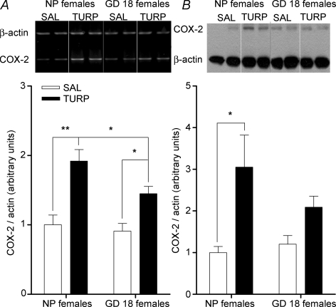
An attenuated fever response to pathogens during late pregnancy is a phenomenon that has been described in several mammalian species, and although mechanisms are not completely understood, decreased prostaglandin E2 (PGE2) synthesis has been implicated. Upstream of PGE2, there is evidence to suggest that anti-inflammatory cytokines such as interleukin-1 receptor antagonist (IL-1ra) could play a significant role. In the present study we addressed the role of pro-inflammatory cytokines during late pregnancy, specifically interleukin-6 (IL-6), an important circulating mediator in fever. Turpentine oil (TURP), a very potent pyrogen and activator of IL-6, was injected into the hind-limb muscle of rats at the 18th day of pregnancy (GD 18) or in non-pregnant (NP) age-matched female controls. As expected, TURP injection induced a highly significant fever in the NP animals, which peaked 11 h post-injection and lasted for over 24 h. This was accompanied by a significant rise in circulating IL-6 levels, which correlated with changes in PGE2 synthesizing enzymes expression in the hypothalamus. In complete contrast, TURP-induced fever was totally absent in GD 18 animals whose body temperature did not deviate from basal values. The lack of response was additionally reflected by the absence of change in IL-6 concentration and by the significant attenuation of PGE2 synthesizing enzymes expression, which correlated with the suppressed expression of SOCS3, a hypothalamic marker of IL-6 activity. Contrary to the changes in circulating IL-6 levels at GD 18, IL-1ra was induced to levels comparable to those of NP females, suggesting that the influence of this anti-inflammatory cytokine on the fever response to TURP is at best minimal. These data further confirm the importance of IL-6 in fever generation and provide evidence that it may be a key component of the attenuated fever response in late pregnancy.


Figures

Figure 1
Figure 1
TURP-induced fever is attenuated in late pregnancy Injection of TURP (100 μl; i.m.) induced a significant rise in core body temperature in NP females (Tc,°C). Animals in the 18th day of pregnancy (GD 18) were completely resistant to the febrile effects of TURP. The arrow indicates the time of injection (animals were injected between 08:00 and 09:00 h). Inset, fever indexes (or area under the curve, AUC) were calculated for each group. AUC in the NP females injected with TURP was significantly greater than both SAL-injected NP females and TURP-injected GD 18 dams. NP females versus SAL-injected NP females: **P < 0.01 and versus TURP-injected GD 18 females: *P < 0.05.
Figure 2
Figure 2
Circulating IL-6 and IL-1ra levels after TURP injection are differentially modulated in late pregnant dams A, 11 h after the injection of TURP (100 μl; i.m.), IL-6 concentration was significantly increased in NP females; this increase was not evident in late-pregnant dams (GD 18), whose levels did not differed from those found in the corresponding SAL-injected controls. TURP-injected NP females versus SAL-injected NP females: **P < 0.01 and versus TURP-injected GD 18 females: *P < 0.05. B, top panel, correlation analysis between plasma IL-6 levels (x axis) and temperature 11 h after injection (y axis) from NP females revealed a significant relationship between these parameters (n = 12 including SAL- and TURP-injected animals; r2= 0.74, P = 0.0003). Bottom panel, the same analysis showed that in GD 18 dams this correlation was not evident (n = 11; r2= 0.08, P = 0.07). C, IL-1ra concentration was significantly increased in both NP and GD 18 females after TURP injection; there was no significant difference in the induced levels of the anti-inflammatory cytokine between these two groups. TURP-injected versus SAL-injected NP females: **P < 0.01; TURP-injected versus SAL-injected GD 18 females: *P < 0.05.
Figure 3
Figure 3
Hypothalamic SOCS3 mRNA, a marker of IL-6 activity, is not induced after TURP challenge during late pregnancy A, in NP females, SOCS3 mRNA expression in the hypothalamus was significantly induced 11 h after TURP injection (100 μl; i.m.); in contrast, this increase was blunted in GD 18 dams. TURP-injected NP females versus SAL-injected NP females: **P < 0.01 and versus TURP-injected GD 18 females: *P < 0.05. Each band in the gel represents one animal; two animals per group are shown. B, top panel, correlation analysis between hypothalamic SOCS3 mRNA levels (x axis) and temperature 11 h after injection (y axis) from NP females revealed a significant relationship between these parameters (n = 12 including SAL- and TURP-injected animals; r2= 0.55, P = 0.0059). Bottom panel, the same analysis showed that in GD 18 dams this correlation was not evident (n = 11; r2= 0.0001, P = 0.89). C, top panel, in NP females, correlation analysis as in B for plasma IL-6 levels (x axis) and hypothalamic SOCS3 mRNA levels (y axis) revealed a significant link between these parameters (n = 12 including SAL- and TURP-injected animals; r2= 0.53, P = 0.0074). Bottom panel, in contrast, in GD 18 dams this correlation (IL-6 versus hypothalamic SOCS3 mRNA) was not evident (n = 11; r2= 0.14, P = 0.25).
Figure 4
Figure 4
COX-2 expression, at the level of mRNA and protein, is differentially regulated in NP and GD 18 females after TURP injection A, TURP injection (100 μl; i.m.) induced a significant increase of COX-2 mRNA expression in the hypothalamus; this induction was reduced, but still present, in GD 18 dams. TURP-injected NP females versus SAL-injected NP females: **P < 0.01; TURP-injected GD 18 females versus SAL-injected GD 18 females and versus TURP-injected NP females: *P < 0.05. Each band in the gel represents one animal; two animals per group are being shown. B, COX-2 protein levels (measured by Western blot) in the hypothalamus of NP females were increased 11 h after TURP injection (100 μl; i.m.); this increase was not present in GD 18 dams. TURP-injected NP females versus SAL-injected NP females: *P < 0.05. Each band in the gel represents one animal; two animals per group are shown.
Figure 5
Figure 5
Attenuated mPGES-1 mRNA expression in the hypothalamus of GD 18 females RT-PCR analysis of mPGES-1 mRNA levels in the hypothalamus revealed a significant induction in NP females after TURP injection (100 μl; i.m.); in comparison, levels of this mRNA were increased to a lesser extent in GD 18 dams. TURP-injected NP females versus SAL-injected NP females: **P < 0.01; TURP-injected GD 18 females versus SAL-injected GD 18 females and versus TURP-injected NP females: *P < 0.05. Each band in the gel represents one animal; two animals per group are being shown.
Figure 6
Figure 6
Hypothalamic IL-1β mRNA induction after TURP is attenuated GD 18-females A, similar to other central markers of inflammation, IL-1β mRNA expression increased in the hypothalamus of NP females, 11 h after TURP injection (100 μl; i.m.). In TURP-injected GD 18 dams the levels of this mRNA were lower, but still significantly induced. TURP-injected NP females versus SAL-injected NP females: **P < 0.01; TURP-injected GD 18 females versus SAL-injected GD 18 females and versus TURP-injected NP females: *P < 0.05. Each band in the gel represents one animal; two animals per group are being shown. B, top panel, hypothalamic IL-1β mRNA levels (x axis) correlated significantly with temperature 11 h after TURP injection (y axis) in NP females (n = 12 including SAL- and TURP-injected animals; r2= 0.77, P = 0.0002). Bottom panel, the same analysis showed that no such correlation existed in GD 18 dams (n = 11; r2= 0.16, P = 0.22). C, top panel, as in B, in NP females hypothalamic IL-1β mRNA levels (y axis) showed a low, but significant correlation with plasma IL-6 levels (x axis) (n = 12 including SAL- and TURP-injected animals; r2= 0.45, P = 0.017). Bottom panel, in GD 18 dams this correlation was still present (n = 11; r2= 0.46, P = 0.046). The expression of microsomal PGE synthase-1 is attenuated in GD 18 dams after TURP injection.

References

    1. Ashdown H, Dumont Y, Ng M, Poole S, Boksa P, Luheshi GN. The role of cytokines in mediating effects of prenatal infection on the fetus: implications for schizophrenia. Mol Psychiatry. 2006a;11:47–55. - PubMed
    1. Ashdown H, Poole S, Boksa P, Luheshi GN. Interleukin-1 receptor antagonist as a modulator of gender differences in the febrile response to lipopolysaccharide in rats. Am J Physiol Regul Integr Comp Physiol. 2006b;292:R1667–R1674. - PubMed
    1. Beagley KW, Gockel CM. Regulation of innate and adaptive immunity by the female sex hormones oestradiol and progesterone. FEMS Immunol Med Microbiol. 2003;38:13–22. - PubMed
    1. Begg DP, Kent S, McKinley MJ, Mathai ML. Suppression of endotoxin-induced fever in near-term pregnant rats is mediated by brain nitric oxide. Am J Physiol Regul Integr Comp Physiol. 2007;292:R2174–R2178. - PubMed
    1. Boksa P. Animal models of obstetric complications in relation to schizophrenia. Brain Res Brain Res Rev. 2004;45:1–17. - PubMed

Publication types

MeSH terms