

Review
doi: 10.1104/pp.107.096156.
Legume evolution: where do nodules and mycorrhizas fit in?
Affiliations
- PMID: 17556520
- PMCID: PMC1914177
- DOI: 10.1104/pp.107.096156
Item in Clipboard
Review
Legume evolution: where do nodules and mycorrhizas fit in?
Plant Physiol.
2007 Jun.
No abstract available
Figures
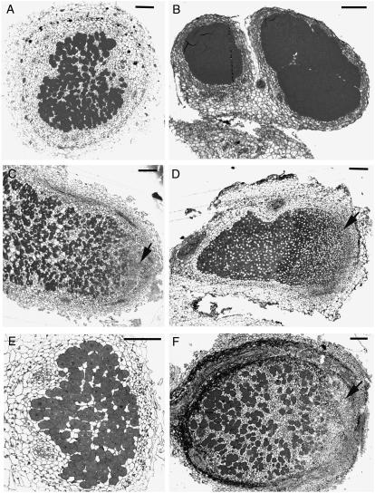
Structure of the main types of legume nodules. A, Sesbania macrantha root nodule. Although morphologically similar to the aeschynomenoid type of nodule seen in B, the infected tissue contains uninfected cells and bacteria are transmitted to infected cells by ITs. B, Aeschynomene rostrata stem nodule. This is typical of a clade of dalbergioid legumes. ITs are never formed and infected tissue contains no uninfected cells. Infection occurs through breaks where lateral or adventitious root initials protrude and a few infected cells divide repeatedly. C, Mimosa himalayana. This structure is typical of all mimosoid and many papilionoid nodules and in most cases follows from root hair infection. There is a clear apical meristem (arrow), and the infected tissue contains a mixture of infected and uninfected cells. ITs convey bacteria to cells newly formed by the meristem. D, Cytisus garden hybrid, typical of many genistoid legumes. ITs are never seen and infected tissue contains no uninfected cells. There is a distinct apical meristem (arrow), which may divide, forming branched nodules or in some cases encircle the root (Lupinus, Lotononis). E, L. uliginosus, a typical determinate nodule as found in many members of tribe Loteae and in phaseoloid legumes such as soybean (Glycine max). Meristematic activity is short lived, infection is via root hairs, and infected tissue contains uninfected cells. F, Erythrophleum ivorense, a typical caesalpinioid nodule with a blunt apex, a clear apical meristem (arrow), and uninfected cells in the infected tissue. Infected cells retain bacteria in modified ITs, known as fixation threads. They may branch repeatedly and be lignified in the outer layers.

A tentative scheme for the evolution of different types of nodule structure. Dashed line, Pathway not fully demonstrated.
References
-
- Alexander IJ (1989) Systematics and ecology of ectomycorrhizal legumes. In CH Stirton, JL Zarucchi, eds, Advances in Legume Biology. Monographs in Systematic Botany, Vol 29. Missouri Botanical Garden, St. Louis, pp 617–624
-
- Allen ON, Allen EK (1981) The Leguminosae: A Source Book of Characteristics, Uses and Nodulation. University of Wisconsin Press, Madison, WI/Macmillan Publishing, London
-
- Batut J, Andersson GE, O'Callaghan DO (2004) The evolution of chronic infection strategies in the α-proteobacteria. Nat Rev Microbiol 2 933–945 - PubMed
-
- Blauenfeldt J, Pa J, Gresshoff PM, Caetano-Anolles G (1994) Nodulation of white clover (Trifolium repens) in the absence of Rhizobium. Protoplasma 179 106–110
Publication types
MeSH terms
LinkOut - more resources
Full Text Sources
Other Literature Sources
