. 2007 Jun 19;104(25):10661-6.
doi: 10.1073/pnas.0610849104. Epub 2007 Jun 7.

Stimulation of subterritories of the subthalamic nucleus reveals its role in the integration of the emotional and motor aspects of behavior

Affiliations

Stimulation of subterritories of the subthalamic nucleus reveals its role in the integration of the emotional and motor aspects of behavior

Luc Mallet et al. Proc Natl Acad Sci U S A. .

Abstract

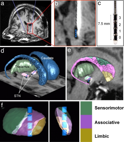
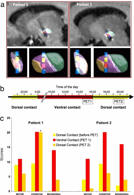
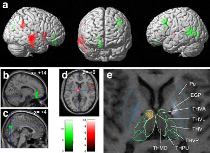
Two parkinsonian patients who experienced transient hypomanic states when the subthalamic nucleus (STN) was stimulated during postoperative adjustment of the electrical parameters for antiparkinsonian therapy agreed to have the mood disorder reproduced, in conjunction with motor, cognitive, and behavioral evaluations and concomitant functional neuroimaging. During the experiment, STN stimulation again induced a hypomanic state concomitant with activation of cortical and thalamic regions known to process limbic and associative information. This observation suggests that the STN plays a role in the control of a complex behavior that includes emotional as well as cognitive and motor components. The localization of the four contacts of the quadripolar electrode was determined precisely with an interactive brain atlas. The results showed that (i) the hypomanic state was caused only by stimulation through one contact localized in the anteromedial STN; (ii) both this contact and the contact immediately dorsal to it improved the parkinsonian motor state; (iii) the most dorsal and ventral contacts, located at the boundaries of the STN, neither induced the behavioral disorder nor improved motor performance. Detailed analysis of these data led us to consider a model in which the three functional modalities, emotional, cognitive, and motor, are not processed in a segregated manner but can be subtly combined in the small volume of the STN. This nucleus would thus serve as a nexus that integrates the motor, cognitive, and emotional components of behavior and might consequently be an effective target for the treatment of behavioral disorders that combine emotional, cognitive, and motor impairment.


Conflict of interest statement

The authors declare no conflict of interest.

Figures

Fig. 1.
Fig. 1.
Method for localizing electrodes implanted in the brain of a patient with Parkinson's disease for stimulation of the STN. (a) Postoperative MRI of a patient showing the electrodes in blue. (b) Identification of the four contacts (blue cylinders) in the electrode MRI artifact. The MRI was reformatted along the trajectory of each electrode and a scaled template of the contacts (1.5 × 1.27 mm cylinder) was positioned at the center of the artifact (20). (c) Medtronic 3389 electrode. (d and e) The three dimensional histological atlas of the basal ganglia is fitted to the patient MRI as described in Experimental Procedures (21). (d) 3D view of caudate, thalamus, and STN after registration with the atlas. (e) Sagittal view of the fitted atlas image including sensorimotor, associative and limbic territories of caudate and STN. (f) Electrode contacts and STN territories after rotation along STN main axes.
Fig. 2.
Fig. 2.
Motor, cognitive and behavioral status of two parkinsonian patients undergoing stimulation of the STN through a ventral contact that induces hypomania and a dorsal contact that ameliorates motor performance. (a) 3D views of the STN territories and contacts for patients 1 and 2 in sagittal views with MRI and after rotation along main STN axes. Therapeutic dorsal contact, yellow; ventral hypomania-inducing contact, red. (b) Schematic representation of the protocol for evaluating cerebral blood flow by PET. Yellow bars represent the normal state during dorsal contact stimulation, red bars the hypomanic state induced by ventral contact stimulation. (c) Motor, neuropsychological and behavioral scores the day before and during the PET protocol. Motor performance: UPDRS III (28) score indicating the severity of parkinsonism. Cognitive abilities: CPT-II score of attention (values >11 are indicative of problems) (24). Behavior: YMRS score (values >12 are considered pathological) (23). The baseline score for patient 1 before PET, which was zero, coincides with the base of the histograms. The CPT-II score returned to baseline values a few hours after the second series of PET acquisitions.
Fig. 3.
Fig. 3.
Brain regions showing activation (red) or deactivation (green) during hypomania induced by stimulation of the STN in patients with Parkinson's disease. (a) Statistical parametric contrast t-maps showing clusters of voxels in which rCBF differed significantly in hypomanic and baseline states. (b and c) Parasagittal sections superimposed on significantly deactivated clusters in deeper brain regions (b, anterior cingulate/subgenual medial prefrontal cortex; c, cuneus). (d) Activated cluster in the left thalamus on an axial section. (e) Superimposition of MRI, single-subject SPM{t} map of patient 1 on the patient-fitted histological atlas showing that the left thalamic activation was centered on the THVA. Similar data were obtained for patient 2. Pu, putamen; GPe, external globus pallidus; THVA, ventral anterior thalamic nucleus; THVL, ventral lateral thalamic nucleus; THVI, ventral intermediate thalamic nucleus; THVP, ventral posterior thalamic nucleus; THPU, pulvinar; THMD, dorsal medial thalamic nucleus.
Fig. 4.
Fig. 4.
Mechanisms by which stimulation of the ventral contact (red cylinder) but not the dorsal contact (yellow cylinder) could induce a hypomanic state. (a–c) The STN is represented along its longest axis with its three functional territories, sensorimotor, associative, and limbic, color coded as in Fig. 1. Green circles are superimposed to indicate different possible zones of diffusion of the stimulating current from the dorsal and ventral contacts: in a radius of 4 mm to structures outside the STN (a), to different STN territories within a radius of 2 mm (b), to the associative territory of the STN within a radius of 1 mm (c). (d) Schematic illustration of the convergence of projections from the cerebral cortex (CX), caudate nucleus (CD), and globus pallidus (GP) on the STN. (e) Schematic illustration, based on data from refs. and –, showing the gradient organization of the different territories of the STN and the disposition of the dendrites of individual neurons which extend over neighboring territories.

Similar articles

Cited by

References

    1. Dalgleish T. Nat Rev Neurosci. 2004;5:583–589. - PubMed
    1. Alexander GE, DeLong MR, Strick PL. Annu Rev Neurosci. 1986;9:357–381. - PubMed
    1. Parent A, Hazrati LN. Brain Res Brain Res Rev. 1995;20:91–127. - PubMed
    1. Bartels A, Zeki S. NeuroReport. 2000;11:3829–3834. - PubMed
    1. Bhatia KP, Marsden CD. Brain. 1994;117:859–876. - PubMed

Publication types

LinkOut - more resources