. 2007 Jul;117(7):1844-55.
doi: 10.1172/JCI30269.

Radiation and inhibition of angiogenesis by canstatin synergize to induce HIF-1alpha-mediated tumor apoptotic switch

Affiliations

Radiation and inhibition of angiogenesis by canstatin synergize to induce HIF-1alpha-mediated tumor apoptotic switch

Claire Magnon et al. J Clin Invest. 2007 Jul.

Abstract

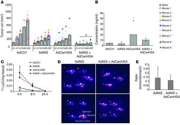
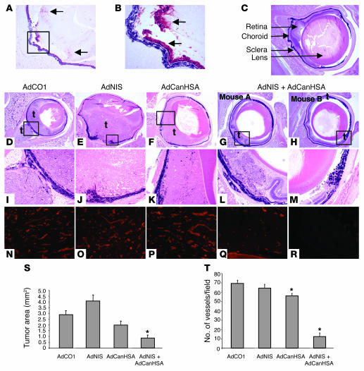
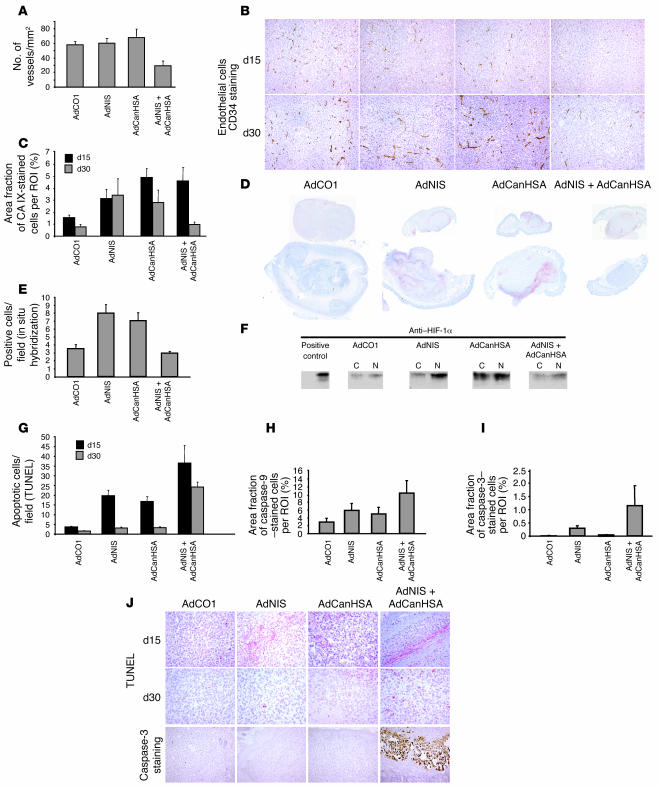
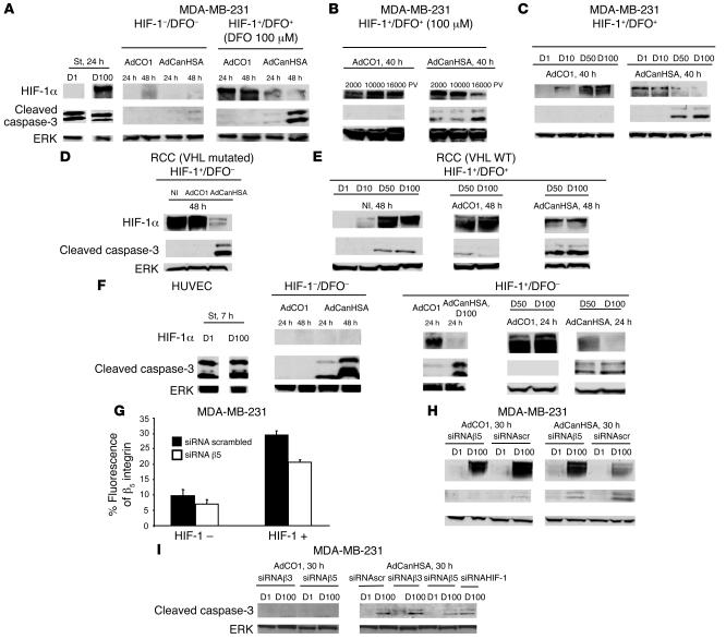
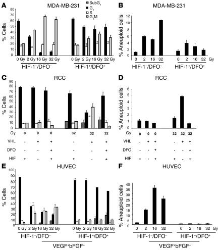
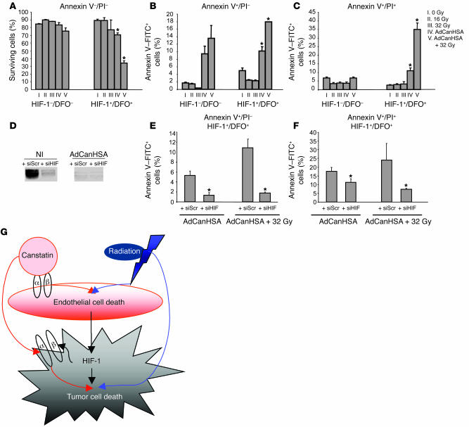
Tumor radioresponsiveness depends on endothelial cell death, which leads in turn to tumor hypoxia. Radiation-induced hypoxia was recently shown to trigger tumor radioresistance by activating angiogenesis through hypoxia-inducible factor 1-regulated (HIF-1-regulated) cytokines. We show here that combining targeted radioiodide therapy with angiogenic inhibitors, such as canstatin, enhances direct tumor cell apoptosis, thereby overcoming radio-induced HIF-1-dependent tumor survival pathways in vitro and in vivo. We found that following dual therapy, HIF-1alpha increases the activity of the canstatin-induced alpha(v)beta(5) signaling tumor apoptotic pathway and concomitantly abrogates mitotic checkpoint and tetraploidy triggered by radiation. Apoptosis in conjunction with mitotic catastrophe leads to lethal tumor damage. We discovered that HIF-1 displays a radiosensitizing activity that is highly dependent on treatment modalities by regulating key apoptotic molecular pathways. Our findings therefore support a crucial role for angiogenesis inhibitors in shifting the fate of radiation-induced HIF-1alpha activity from hypoxia-induced tumor radioresistance to hypoxia-induced tumor apoptosis. This study provides a basis for developing new biology-based clinically relevant strategies to improve the efficacy of radiation oncology, using HIF-1 as an ally for cancer therapy.


Figures

Figure 1
Figure 1. Therapeutic efficiency of AdNIS-131I therapy combined with AdCanHSA in vivo.
(A) Growth of AdCO1-, AdNIS-, AdCanHSA-, and AdNIS-AdCanHSA–infected MDA-MB-231 xenografted tumors (2 intratumor injections of the appropriate adenoviruses, 72 hours apart) on nu/nu mice after injection of 300 μCi of 131I. Tumor volumes were measured once a week over 4 weeks. Results are mean ± SEM for 9 mice for each treatment group. Representative of 4 separate experiments. *P < 0.05. (B) ELISA quantification of CanHSA in the sera of the same mice represented above, at day 4 after injection of iodide. (C) In vivo kinetics of iodide uptake in AdCO1-, AdNIS-, AdCanHSA-, and AdCanHSA-AdNIS–infected MDA-MB-231 tumors (n = 3 per group). Infected tumors were removed 4, 8, and 24 hours following injection of 131I (300 μCi) i.p. 131I was measured using a gamma counter. Results are expressed as the mean number of μCi per milligram of tumor tissue ± SEM. (D) In vivo imaging of mice harboring AdNIS- or AdCanHSA-AdNIS–infected MDA-MB-231 xenografts (left flank) 4 hours after i.p. injection of 50 μCi of 123I and 24 hours before 131I injection. Images were acquired through scintigraphy over a 10-minute exposure and have an equivalent background. There was also physiological accumulation of iodide in the bladder, stomach, and thyroid gland. Mice were treated with l-thyroxine to avoid massive uptake by the thyroid. (E) In vivo assessment of iodide uptake by AdNIS- or AdCanHSA-AdNIS–infected MDA-MB-231 tumors 4 hours after 123I injection, as described for D. A fixed region of interest (ROI) was drawn and was imposed on each tumor and on each stomach (as standard uptake). Pixel intensity per ROI was measured with ImageJ software. Results are expressed as the ratio (tumor/stomach) between the maximal pixel intensity in the 2 ROIs.
Figure 2
Figure 2. Combined effects of AdNIS-131I therapy with AdCanHSA in transgenic TRP-1 mouse model.
(A) Immunohistological analysis of AdNIS-infected RPE tumor with an anti-NIS polyclonal antibody (original magnification, ×50). (B) Higher-magnification of boxed area in A (original magnification, ×200). Positive cells were located in the RPE tumor and in the retina (arrows). (C) Representative nontumor eye section with location of retina, choroid, sclera, and lens (arrows). (DH) Histology of the RPE tumor 45 days after birth. Representative transgenic eye section treated with 2 systemic injections of Adx and a single intraorbital injection of Ady, and a single 131I injection of 300 μCi. AdCO1 and AdCO1 (D); AdCO1 and AdNIS (E); AdCanHSA and AdCO1 (F); or AdCanHSA and AdNIS (G and H) (original magnification, ×25). Note that AdCO1-treated tumor cells grew consistently and occupied half of the eyeball when mice were 45 days old (D). In contrast, only a few tumor cells persisted in AdCanHSA-AdNIS–treated eyeball (H). t, tumor. (IM) Higher-magnification images of areas in black boxes in DH, respectively (original magnification, ×100). (S) Measurement of tumor areas in the posterior eyeball of each TRP-1 transgenic mouse treated with the appropriate adenoviruses. Results are the mean ± SEM (n = 10). (NR) Assessment of intratumor vascularization using lectin immunostaining after each treatment described above (original magnification, ×100). AdCO1 and AdCO1 (N); AdCO1 and AdNIS (O); AdCanHSA and AdCO1 (P); AdCanHSA and AdNIS (Q and R) (same as in black boxes in DH). Original magnification, ×200. (T) Mean number of intratumor vessels for each group ± SEM. *P < 0.05.
Figure 3
Figure 3. In vivo, detection of hypoxic, apoptotic, and endothelial markers in MDA-MB-231 xenografted tumors following AdNIS-131I therapy combined with AdCanHSA.
(A) The number of CD34-stained intratumor microvessels per square millimeter was quantified by digital analysis of tumor tissue sections at day 30 after infection. Original magnification, ×100. (B) Representative images of CD34 endothelial immunostaining. (C) Quantification of the area fraction of hypoxic CAIX-stained cells within digitized sections from AdCO1-, AdNIS-, AdCanHSA-, and AdCanHSA-AdNIS–injected tumors at days 15 and 30 (i.e., at days 8 and 20 after injection of 131I). (D) Representative CAIX-stained digitized sections for each group. (E) Quantification of HIF-1 mRNA levels by in situ hybridization analysis. The number of cells per field producing mRNA HIF-1α was quantified within tumors sections (×40) at day 30 following injections of appropriate adenoviruses listed above. (F) Western blotting analysis of HIF-1α protein levels within nuclear (N) and cytoplasmic (C) extracts of adenovirus-infected xenografted tumors. A whole-cell lysate of RCC (VHL mutated) cells was considered as positive control. Note that HIF-1α and CAIX have a similar expression pattern at day 30. (G) The proportion of apoptotic cells per field was quantified within sections (×200) (TUNEL method) at days 15 and 30 after infection. Quantification of the area fraction of caspase-9– (H) and caspase-3–stained cells (I) within digitized sections from treated tumors at day 30 as described in Figure 1A. (J) Representative illustrations of both TUNEL and caspase-3 staining. Columns, mean ± SEM of 9 mice for each treated group. All digitized paraffin-embedded tumor sections were obtained from the experiment described in Figure 1A. Original magnification, ×200.
Figure 4
Figure 4. HIF-1 is required for potentiation of integrin-mediated tumor apoptotic signaling pathway induced by CanHSA.
The expression levels of both HIF-1α and caspase-3 were assessed by Western blotting of whole-cell extracts prepared from AdCO1- or AdCanHSA-infected MDA-MB-231 cells, in the presence or absence of DFO (A) at different time points following infection (B) with several MOIs (2,000, 10,000, or 16,000 particle viral [PV]) or (C) at different concentrations of DFO (1, 10, 50, and 100 μM [D1, D10, D50, and D100]). Immunoblots of whole-cell extracts, from noninfected (NI) or AdCO1- or AdCanHSA-infected RCC cells expressing VHL mutated (D) or VHL WT (E) in the presence or absence of DFO, confirmed that HIF-1 expression potentiates caspase-3 cleavage following infection with AdCanHSA. (F) Western blotting of protein extracts from HUVECs showed that HIF-1α is not required to induce the CanHSA-mediated apoptotic process in endothelial cells. Note that HIF-1α expression slightly accelerated caspase-3 cleavage. Staurosporine (St), a known inducer of caspase-dependent apoptosis, was used as a positive control. To determine whether αvβ5 integrins promote the CanHSA-induced HIF-1α signaling tumor apoptotic pathway, expression of αvβ5 was silenced through siRNA β5 on MDA-MB-231 cells expressing or not expressing HIF-1α (FACS analysis as previously described; ref. 27) (G) and Western blotting was done on whole-cell extracts from AdCO1- or AdCanHSA-infected MDA-MB-231 cells, which were transfected with random siRNA (siRNA scrambled) or siRNA β5 (H). (I) In parallel, to investigate whether αvβ3 integrins take part in the HIF-1–dependent tumor apoptotic process, MDA-MB-231 cells were also transfected with siRNA β3 and then infected with AdCO1 and AdCanHSA. Each experiment was done twice. To confirm whether CanHSA triggers an HIF-1–dependent apoptotic process, we transfected an siRNA HIF-1 within the MDA-MB-231 cells.
Figure 5
Figure 5. HIF-1 mediates the control of mitosis checkpoint, aneuploidy, and haploploidy following radiation.
Cell-cycle distribution of cells exposed to escalating doses of γ-radiation (2, 16, and 32 Gy, for each dose, twice, 3 hours apart). Percentage of cells of the population found in G1, S, G2/M phase and tetraploidy for MDA-MB-231 cells (A and B), RCC cells (VHL mutated or WT) (C and D), and HUVECs (E and F). Bars indicate SEM. Each experiment was done twice. All experiments were performed with VEGF and bFGF, confirming that HIF-1 controls this checkpoint in a growth factor–independent manner.
Figure 6
Figure 6. HIF-1 is the key regulator of the tumor apoptotic switch following combined therapy.
FACS, annexin V–FITC, and propidium iodide [PI] profiles of MDA-MB-231 cells that were viable (A); in early apoptosis with membrane integrity (B); or in late apoptosis with death following AdCanHSA infection combined or not with γ-radiation (C). (D) To assess the suppression of HIF-1α expression, Western blotting was done on noninfected or AdCanHSA-infected MDA-MB-231 cells that were transfected with random siRNA (siRNA scrambled [siScr]) or siRNA HIF-1α (siHIF). (E and F) Annexin V–FITC staining was performed on transfected MDA-MB-231 cells following AdCanHSA infection combined or not with γ-radiation. Bars represent SEM. Each experiment was done twice. *P < 0.05. (G) Schematic illustration of the role of HIF-1 in tumor response to canstatin and/or radiation. Endothelial cell death is directly triggered by both canstatin through an integrin-mediated mitochondrial apoptotic mechanism and radiation exposures through an apoptotic pathway. The resulting microvascular dysfunction induces HIF-1α expression, as a mandatory signal regulating the tumor adaptive response. HIF-1α in turn upregulates integrin-receptor expression on tumor cell surface, enhancing specifically HIF-1α signaling tumor apoptotic pathway triggered by canstatin. Moreover, HIF-1 confers the conversion of sublethal radiation damage, such as mitotic arrest and tetraploid status, into lethal lesions through aberrant mitosis in both endothelial and tumor cells. Collectively, induction of profound tumor cell death requires both canstatin-induced tumor apoptotic pathway and radiation-mediated mitotic catastrophe through an HIF-1 signaling molecular mechanism.

References

    1. Schlumberger M. Papillary and follicular thyroid carcinoma. N. Engl. J. Med. 1998;338:297–306. - PubMed
    1. Cooper D.S., et al. Management guidelines for patients with thyroid nodules and differentiated thyroid cancer. Thyroid. 2006;16:109–142. - PubMed
    1. Dai G., Levy O., Carrasco N. Cloning and characterization of the thyroid iodide symporter. Nature. 1996;379:458–460. - PubMed
    1. Eskandari S., et al. Thyroid Na+/I– symporter. Mechanism, stoichiometry, and specificity. J. Biol. Chem. . 1997;272:27230–27238. - PubMed
    1. Filetti S., et al. Sodium/iodide symporter: a key transport system in thyroid cancer cell metabolism. Eur. J. Endocrinol. 1999;141:443–457. - PubMed

Publication types

MeSH terms