

Modulation of cortical oscillatory activity during transcranial magnetic stimulation
- PMID: 17557296
- PMCID: PMC6870908
- DOI: 10.1002/hbm.20423
Modulation of cortical oscillatory activity during transcranial magnetic stimulation
Abstract
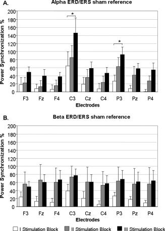
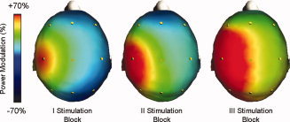
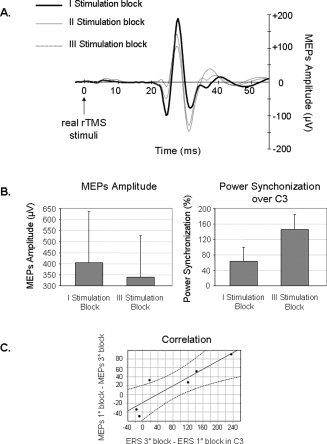
Transcranial magnetic stimulation (TMS) can transiently modulate cortical excitability, with a net effect depending on the stimulation frequency (< or =1 Hz inhibition vs. > or =5 Hz facilitation, at least for the motor cortex). This possibility has generated interest in experiments aiming to improve deficits in clinical settings, as well as deficits in the cognitive domain. The aim of the present study was to investigate the on-line effects of low frequency (1 Hz) TMS on the EEG oscillatory activity in the healthy human brain, focusing particularly on the outcome of these modulatory effects in relation to the duration of the TMS stimulation. To this end, we used the event-related desynchronization/synchronization (ERD/ERS) approach to determine the patterns of oscillatory activity during two consecutive trains of sham and real TMS. Each train of stimulation was delivered to the left primary motor cortex (MI) of healthy subjects over a period of 10 min, while EEG rhythms were simultaneously recorded. Results indicated that TMS induced an increase in the power of brain rhythms that was related to the period of the stimulation, i.e. the synchronization of the alpha band increased with the duration of the stimulation, and this increase was inversely correlated with motor-evoked potentials (MEPs) amplitude. In conclusion, low frequency TMS over primary motor cortex induces a synchronization of the background oscillatory activity on the stimulated region. This induced modulation in brain oscillations seems to increase coherently with the duration of stimulation, suggesting that TMS effects may involve short-term modification of the neural circuitry sustaining MEPs characteristics.
(Copyright) 2006 Wiley-Liss, Inc.
Figures
Similar articles
-
Modulation of cortical oscillatory activities induced by varying single-pulse transcranial magnetic stimulation intensity over the left primary motor area: a combined EEG and TMS study.Neuroimage. 2005 Oct 1;27(4):896-908. doi: 10.1016/j.neuroimage.2005.05.013. Neuroimage. 2005. PMID: 16054397 Clinical Trial.
-
Now I am ready-now i am not: The influence of pre-TMS oscillations and corticomuscular coherence on motor-evoked potentials.Cereb Cortex. 2014 Jul;24(7):1708-19. doi: 10.1093/cercor/bht024. Epub 2013 Feb 8. Cereb Cortex. 2014. PMID: 23395847
-
The impact of GABAergic drugs on TMS-induced brain oscillations in human motor cortex.Neuroimage. 2017 Dec;163:1-12. doi: 10.1016/j.neuroimage.2017.09.023. Epub 2017 Sep 14. Neuroimage. 2017. PMID: 28917695 Clinical Trial.
-
Measuring Brain Stimulation Induced Changes in Cortical Properties Using TMS-EEG.Brain Stimul. 2015 Nov-Dec;8(6):1010-20. doi: 10.1016/j.brs.2015.07.029. Epub 2015 Jul 17. Brain Stimul. 2015. PMID: 26275346 Review.
-
Transcranial magnetic stimulation.Handb Clin Neurol. 2019;160:559-580. doi: 10.1016/B978-0-444-64032-1.00037-0. Handb Clin Neurol. 2019. PMID: 31277876 Review.
Cited by
-
Assessing cortical network properties using TMS-EEG.Hum Brain Mapp. 2013 Jul;34(7):1652-69. doi: 10.1002/hbm.22016. Epub 2012 Feb 29. Hum Brain Mapp. 2013. PMID: 22378543 Free PMC article.
-
EEG theta and beta bands as brain oscillations for different knee osteoarthritis phenotypes according to disease severity.Sci Rep. 2022 Jan 27;12(1):1480. doi: 10.1038/s41598-022-04957-x. Sci Rep. 2022. PMID: 35087082 Free PMC article.
-
Theta-burst Transcranial Magnetic Stimulation Alters the Functional Topography of the Cortical Motor Network.Malays J Med Sci. 2015 Dec;22(Spec Issue):36-44. Malays J Med Sci. 2015. PMID: 27006636 Free PMC article.
-
Effects of 10 Hz rTMS on the neural efficiency of working memory.J Cogn Neurosci. 2010 Mar;22(3):447-56. doi: 10.1162/jocn.2009.21209. J Cogn Neurosci. 2010. PMID: 19309294 Free PMC article.
-
Disrupted Hippocampal Theta-Gamma Coupling and Spike-Field Coherence Following Experimental Traumatic Brain Injury.bioRxiv [Preprint]. 2024 Sep 12:2024.05.30.596704. doi: 10.1101/2024.05.30.596704. bioRxiv. 2024. PMID: 39314320 Free PMC article. Preprint.
References
-
- Amassian VE,Cracco RQ,Maccabee PJ ( 1989): Focal stimulation of human cerebral cortex with the magnetic coil: A comparison with electrical stimulation. Electroencephalogr Clin Neurophysiol 74: 401–416. - PubMed
-
- Barker AT,Jalinous R,Freeston IL ( 1985): Non‐invasive magnetic stimulation of human motor cortex. Lancet 1: 1106–1107. - PubMed
-
- Barker AT,Freeston IL,Jabinous R,Jarratt JA ( 1986): Clinical evaluation of conduction time measurements in central motor pathways using magnetic stimulation of human brain. Lancet 1: 1325–1326. - PubMed
-
- Berardelli A,Inghilleri M,Rothwell JC,Romeo S,Curra A,Gilio F,Modugno N,Manfredi M ( 1998): Facilitation of muscle evoked responses after repetitive cortical stimulation in man. Exp Brain Res 122: 79–84. - PubMed
-
- Bonato C,Miniussi C,Rossini PM ( 2006): Transcranial magnetic stimulation and cortical evoked potentials: A TMS/EEG co‐registration study. Clin Neurophysiol 117: 1699–1707. - PubMed
Publication types
MeSH terms
LinkOut - more resources
Full Text Sources
Other Literature Sources
