. 2007 Oct 15;93(8):2870-6.
doi: 10.1529/biophysj.107.109728. Epub 2007 Jun 8.

Rows of ATP synthase dimers in native mitochondrial inner membranes

Affiliations

Rows of ATP synthase dimers in native mitochondrial inner membranes

Nikolay Buzhynskyy et al. Biophys J. .

Abstract

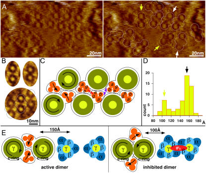
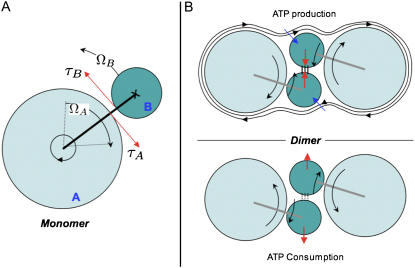
The ATP synthase is a nanometric rotary machine that uses a transmembrane electrochemical gradient to form ATP. The structures of most components of the ATP synthase are known, and their organization has been elucidated. However, the supramolecular assembly of ATP synthases in biological membranes remains unknown. Here we show with submolecular resolution the organization of ATP synthases in the yeast mitochondrial inner membranes. The atomic force microscopy images we have obtained show how these molecules form dimers with characteristic 15 nm distance between the axes of their rotors through stereospecific interactions of the membrane embedded portions of their stators. A different interaction surface is responsible for the formation of rows of dimers. Such an organization elucidates the role of the ATP synthase in mitochondrial morphology. Some dimers have a different morphology with 10 nm stalk-to-stalk distance, in line with ATP synthases that are accessible to IF1 inhibition. Rotation torque compensation within ATP synthase dimers stabilizes the ATP synthase structure, in particular the stator-rotor interaction.


Figures

FIGURE 1
FIGURE 1
Coomassie brilliant blue stained SDS-PAGE of yeast MIM fractions. Bands labeled and marked with solid lines were identified on the basis of liquid chromatography and mass spectrometry analysis and gel migration behavior. Bands marked with dotted lines were not identified.
FIGURE 2
FIGURE 2
Overview AFM analysis of MIM. (A) MIM adsorbed on mica. The folded parts of the membrane are marked with white arrows; the membrane area opened with the AFM tip is marked by the red arrow. (B) Topograph of the flattened membrane area shown in A. The AFM tip contours circular complexes of ∼8 nm in diameter and intercomplex distances of ∼15 nm corresponding to c-rings of ATP synthase. (C and D) Overview images reveal arrays of c-ring complexes densely packed. (E) Row of intercalated dimers. The c-ring rotor appears as a ∼1 nm indentation.
FIGURE 3
FIGURE 3
Supramolecular architecture of ATP synthases. (A) High-resolution AFM topograph of MIM. (Right) ATP synthase dimers are outlined. (B) Average topographies. Left: Dimer with 15 nm stalk-to-stalk distance. (Right) Dimer with 10 nm stalk-to-stalk distance. (Bottom) Outlines indicate the molecular packing along a row of dimers. (C) Schematic representation of dimer and oligomer assembly. The positions of the molecules correspond to the average topograph (B, bottom panel). The assignment of subunits e and g at dimer interfaces and the transmembrane portion of b at interfaces along the dimer row are based on Arnold et al. (12) and Paumard et al. (13). (Left) Black arrows indicate rotor and stator rotation torque direction. (D) Stalk-to-stalk distance histogram (n = 58). Two populations of dimers are distinguished; most dimers have 15 nm stalk-to-stalk distance (white arrows in A); a minor fraction has 10 nm stalk-to-stalk distance (yellow arrows in A). (E) Schematic representation of active (left) and IF1-inhibited (right) ATP synthase dimers. The rotation torques stabilize the dimer during ATP synthesis and separate FO stator portions during ATP hydrolysis.
FIGURE 4
FIGURE 4
Theoretical considerations about the ATP synthase dimer formation. (A) Monomer of ATP synthase; opposite torques τA and τB are generated on the rotor A and stator B. (B) Functional dimers of ATP synthase. Hydrodynamic (blue arrows) and torque (red arrows) forces stabilize the interaction within the ATP synthase dimer during the ATP production, whereas during the consumption the forces tend to destabilize the dimer.

References

    1. Boyer, P. 1997. The ATP synthase—a splendid molecular machine. Annu. Rev. Biochem. 66:717–749. - PubMed
    1. Stock, D., C. Gibbons, I. Arechaga, A. G. Leslie, and J. E. Walker. 2000. The rotary mechanism of ATP synthase. Curr. Opin. Struct. Biol. 10:672–679. - PubMed
    1. Abrahams, J. P., A. G. W. Leslie, R. Lutter, and J. E. Walker. 1994. Structure at 2.8 angstrom resolution of F1-ATPase from bovine heart mitochondria. Nature. 370:621–628. - PubMed
    1. Gibbons, C., M. G. Montgomery, A. G. Leslie, and J. E. Walker. 2000. The structure of the central stalk in bovine F1-ATPase at 2.4 Å resolution. Nat. Struct. Biol. 7:1055–1061. - PubMed
    1. Stock, D., A. G. Leslie, and J. E. Walker. 1999. Molecular architecture of the rotary motor in ATP synthase. Science. 286:1700–1705. - PubMed

Publication types

MeSH terms

Substances

LinkOut - more resources