. 2007 Jun;19(6):1947-63.
doi: 10.1105/tpc.107.051391. Epub 2007 Jun 8.

Structural evidence for the evolution of xyloglucanase activity from xyloglucan endo-transglycosylases: biological implications for cell wall metabolism

Affiliations

Structural evidence for the evolution of xyloglucanase activity from xyloglucan endo-transglycosylases: biological implications for cell wall metabolism

Martin J Baumann et al. Plant Cell. 2007 Jun.

Abstract

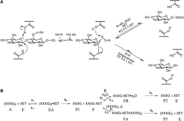
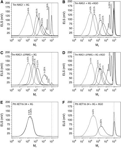
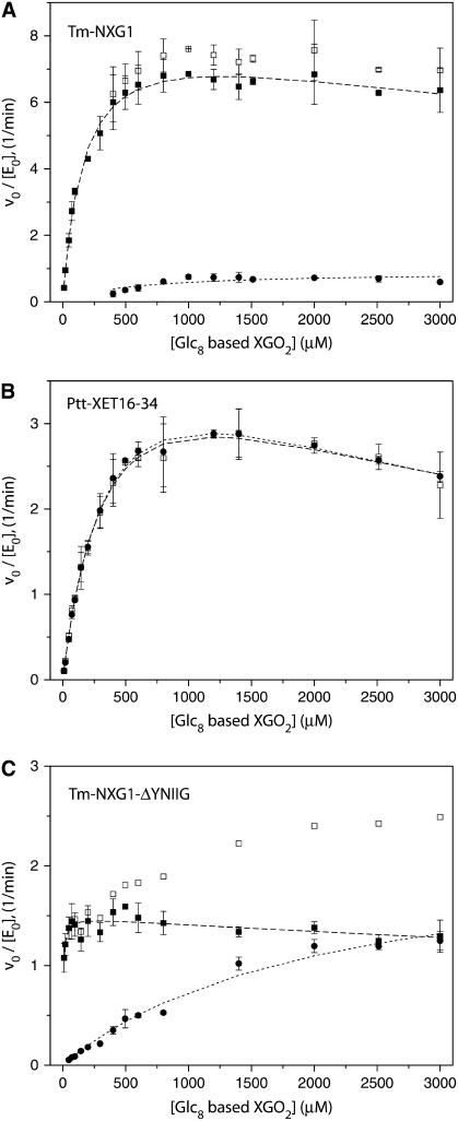
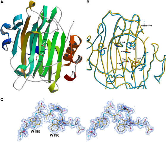
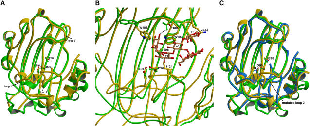
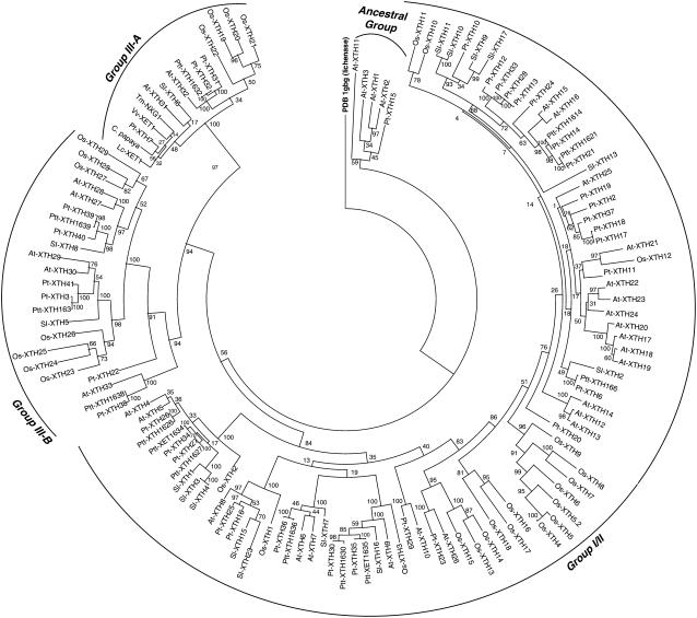
High-resolution, three-dimensional structures of the archetypal glycoside hydrolase family 16 (GH16) endo-xyloglucanases Tm-NXG1 and Tm-NXG2 from nasturtium (Tropaeolum majus) have been solved by x-ray crystallography. Key structural features that modulate the relative rates of substrate hydrolysis to transglycosylation in the GH16 xyloglucan-active enzymes were identified by structure-function studies of the recombinantly expressed enzymes in comparison with data for the strict xyloglucan endo-transglycosylase Ptt-XET16-34 from hybrid aspen (Populus tremula x Populus tremuloides). Production of the loop deletion variant Tm-NXG1-DeltaYNIIG yielded an enzyme that was structurally similar to Ptt-XET16-34 and had a greatly increased transglycosylation:hydrolysis ratio. Comprehensive bioinformatic analyses of XTH gene products, together with detailed kinetic data, strongly suggest that xyloglucanase activity has evolved as a gain of function in an ancestral GH16 XET to meet specific biological requirements during seed germination, fruit ripening, and rapid wall expansion.


Figures

Figure 1.
Figure 1.
The Canonical Retaining Mechanism of Glycosyl Transfer Showing Product Partitioning between Hydrolysis and Transglycosylation. (A) The chemical mechanism. (B) Full kinetic scheme for the minimal xylogluco-oligosaccharide donor substrate XXXGXXXG under steady state conditions in the absence of products. Substrates (A = XXXGXXXG, B = H2O), enzyme intermediates (E, EA, F, FA, and FB), and products (P1 to P3) are denoted with uppercase letters, and individual rate constants (kn) are shown.
Figure 2.
Figure 2.
Size-Exclusion Chromatography of the Products of Tm-NXG1, Tm-NXG1-ΔYNIIG, and Ptt-XET16-34 Operating on Xyloglucan Polysaccharide (Initial Mr 105 to 107) in the Absence and Presence of Xyloglucan Oligosaccharides (Mr 103). Incubation times are indicated above each chromatogram. The vertical dotted line in all panels indicates the elution of Glc4-based XGOs. Protein concentrations were as follows: Tm-NXG1 (9 mg/L), Tm-NXG1-ΔYNIIG (10 mg/L), and Ptt-XET16-34 (10 mg/L). Other experimental conditions are described in Methods.
Figure 3.
Figure 3.
Initial Rate Kinetics as a Function of XGOGlc8 Concentration. Kinetics are shown for Tm-NXG1 (A), Tm-NXG1-ΔYNIIG (B), and Ptt-XET16-34 (C). Closed circles, rate of XGOGlc12 production due to transglycosylation (2 XGOGlc8 → XGOGlc12 + XGOGlc4); open squares, total rate of XGOGlc4 production; closed squares, corrected rate of XGOGlc4 production obtained by subtracting the contribution of XGOGlc4 release due to substrate transglycosylation. This observed rate is twice the actual catalytic rate, according to the stoichiometry of the hydrolysis reaction (XGOGlc8 → 2 XGOGlc4). Error bars indicate sd for triplicate (A), duplicate (B), and duplicate (C) measurements.
Figure 4.
Figure 4.
Three-Dimensional Structures of Tm-NXG1 and Tm-NXG2. (A) Ribbon representation of the crystallographic structure of Tm-NXG1. The polypeptide chain is colored from blue (N terminus) to red (C terminus). The three strictly conserved amino acids forming the catalytic machinery are labeled. (B) Superimposition of the structures of Tm-NXG1 (yellow) and Tm-NXG2 (blue) highlighting the different conformations of the loops that surround the active site groove. The catalytic residues, the three Trp residues that line the substrate binding cleft, and Tyr-122 and Ile-124 are labeled. Due to high disorder, the loop residues between Thr-192 and Lys-196 could not be modeled in the crystal structure of Tm-NXG2. (C) Wall-eyed stereo view of the final 2Fo-Fc electron density map displayed at a 1σ level around the loop containing Trp-185 and Trp-190 in the crystal structure of Tm-NXG1.
Figure 5.
Figure 5.
Structural Comparison of Ptt-XET16-34 and Tm-NXG1. (A) Ribbon representation of the superimposition of Ptt-XET16-34 (green) onto Tm-NXG1 (yellow) illustrating the structural differences that appear mainly in loops 1 through 3 and in the C-terminal region. The catalytic machinery is labeled. (B) Close-up view into the positive subsites of Ptt-XET16-34. The superimposition was performed using the complex structure of Ptt-XET16-34 with bound XLLG (PDB identifier 1UMZ). Asp-178 in Ptt-XET16-34 forms a hydrogen bond to a xylosyl branch of the bound oligosaccharide. Ser-189 in Tm-NXG1 (or Tm-NXG2) is too distant to be able to form an equivalent bond. Gly-183 is replaced by Asn-194 in Tm-NXG1, which would collide with bound sugar in this loop conformation. Ile-124, which is part of the critical loop insertion in Tm-NXG1, would collide with the glucose unit bound in subsite +1 of the acceptor binding cleft. (C) Ribbon representation of the superimposition of Tm-NXG1-ΔYNIIG (blue) onto Ptt-XET16-34 (green) and Tm-NXG1 (yellow) illustrating that the Cα trace of the truncated loop 2 in Tm-NXG1-ΔYNIIG is now closer to that of Ptt-XET16-34. The catalytic machinery is labeled.
Figure 6.
Figure 6.
Unrooted Phylogenetic Tree of ∼130 Full-Length XTH Gene Products and Bacillus licheniformis Lichenase (PDB identifier 1GBG; GenPept Accession Number CAA40547). Bootstrap values from 100 maximum likelihood resamplings are indicated.

References

    1. Aspeborg, H., et al. (2005). Carbohydrate-active enzymes involved in the secondary cell wall biogenesis in hybrid aspen. Plant Physiol. 137 983–997. - PMC - PubMed
    1. Aubert, D., and Herzog, M. (1996). A new cDNA encoding a xyloglucan endo-transglycosylase-related polypeptide (AtXTR8) preferentially expressed in seedling, root and stem of Arabidopsis thaliana. Plant Sci. 121 187–196.
    1. Barbeyron, T., Gerard, A., Potin, P., Henrissat, B., and Kloareg, B. (1998). The kappa-carrageenase of the marine bacterium Cytophaga drobachiensis. Structural and phylogenetic relationships within family-16 glycoside hydrolases. Mol. Biol. Evol. 15 528–537. - PubMed
    1. Becnel, J., Natarajan, M., Kipp, A., and Braam, J. (2006). Developmental expression patterns of Arabidopsis XTH genes reported by transgenes and Genevestigator. Plant Mol. Biol. 61 451–467. - PubMed
    1. Bendtsen, J.D., Nielsen, H., von Heijne, G., and Brunak, S. (2004). Improved prediction of signal peptides: SignalP 3.0. J. Mol. Biol. 340 783–795. - PubMed

Publication types

MeSH terms