

Metal ions may suppress or enhance cellular differentiation in Candida albicans and Candida tropicalis biofilms
- PMID: 17557844
- PMCID: PMC1951024
- DOI: 10.1128/AEM.02711-06
Metal ions may suppress or enhance cellular differentiation in Candida albicans and Candida tropicalis biofilms
Abstract
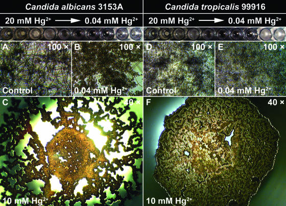
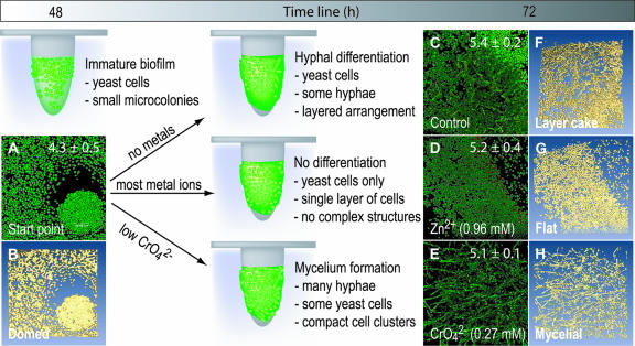
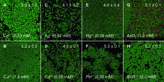
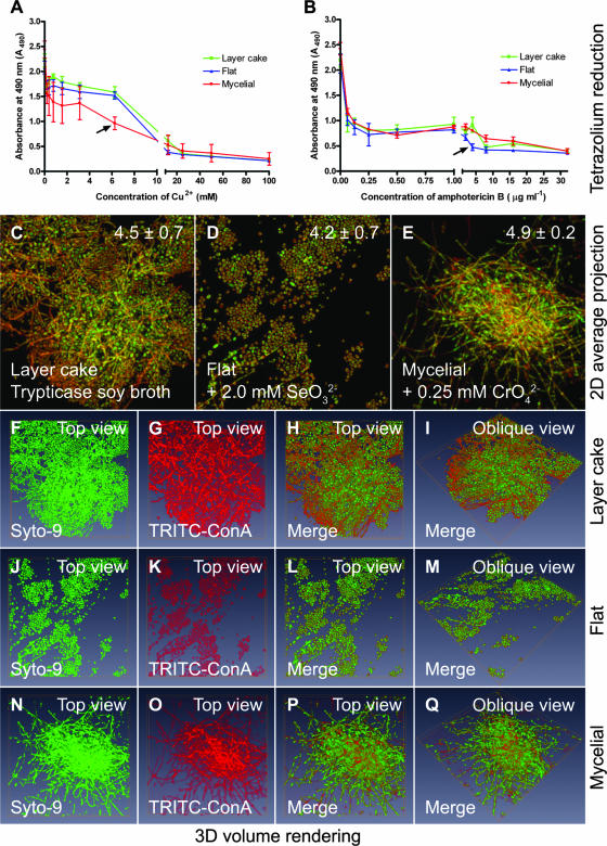
Candida albicans and Candida tropicalis are polymorphic fungi that develop antimicrobial-resistant biofilm communities that are characterized by multiple cell morphotypes. This study investigated cell type interconversion and drug and metal resistance as well as community organization in biofilms of these microorganisms that were exposed to metal ions. To study this, Candida biofilms were grown either in microtiter plates containing gradient arrays of metal ions or in the Calgary Biofilm Device for high-throughput susceptibility testing. Biofilm formation and antifungal resistance were evaluated by viable cell counts, tetrazolium salt reduction, light microscopy, and confocal laser scanning microscopy in conjunction with three-dimensional visualization. We discovered that subinhibitory concentrations of certain metal ions (CrO(4)(2-), Co(2+), Cu(2+), Ag(+), Zn(2+), Cd(2+), Hg(2+), Pb(2+), AsO(2)(-), and SeO(3)(2-)) caused changes in biofilm structure by blocking or eliciting the transition between yeast and hyphal cell types. Four distinct biofilm community structure types were discerned from these data, which were designated "domed," "layer cake," "flat," and "mycelial." This study suggests that Candida biofilm populations may respond to metal ions to form cell-cell and solid-surface-attached assemblages with distinct patterns of cellular differentiation.
Figures

Similar articles
-
A subpopulation of Candida albicans and Candida tropicalis biofilm cells are highly tolerant to chelating agents.FEMS Microbiol Lett. 2007 Jul;272(2):172-81. doi: 10.1111/j.1574-6968.2007.00745.x. Epub 2007 May 8. FEMS Microbiol Lett. 2007. PMID: 17490429
-
Metal resistance in Candida biofilms.FEMS Microbiol Ecol. 2006 Mar;55(3):479-91. doi: 10.1111/j.1574-6941.2005.00045.x. FEMS Microbiol Ecol. 2006. PMID: 16466387
-
In vitro effectiveness of anidulafungin against Candida sp. biofilms.J Antibiot (Tokyo). 2013 Dec;66(12):701-4. doi: 10.1038/ja.2013.83. Epub 2013 Sep 11. J Antibiot (Tokyo). 2013. PMID: 24022607
-
Dispersal of single and mixed non-albicans Candida species biofilms by β-1,3-glucanase in vitro.Microb Pathog. 2017 Dec;113:342-347. doi: 10.1016/j.micpath.2017.10.057. Epub 2017 Nov 1. Microb Pathog. 2017. PMID: 29101060
-
How Biofilms Evade Host Defenses.Microbiol Spectr. 2015 Jun;3(3). doi: 10.1128/microbiolspec.MB-0012-2014. Microbiol Spectr. 2015. PMID: 26185085 Review.
Cited by
-
Evaluation of Biofilm Formation in Candida tropicalis Using a Silicone-Based Platform with Synthetic Urine Medium.Microorganisms. 2020 May 1;8(5):660. doi: 10.3390/microorganisms8050660. Microorganisms. 2020. PMID: 32369936 Free PMC article.
-
Candida Biofilms: Development, Architecture, and Resistance.Microbiol Spectr. 2015 Aug;3(4):10.1128/microbiolspec.MB-0020-2015. doi: 10.1128/microbiolspec.MB-0020-2015. Microbiol Spectr. 2015. PMID: 26350306 Free PMC article. Review.
-
Impact of manganese on biofilm formation and cell morphology of Candida parapsilosis clinical isolates with different biofilm forming abilities.FEMS Yeast Res. 2019 Sep 1;19(6):foz057. doi: 10.1093/femsyr/foz057. FEMS Yeast Res. 2019. PMID: 31403663 Free PMC article.
-
Biofilm Formation in Medically Important Candida Species.J Fungi (Basel). 2023 Sep 22;9(10):955. doi: 10.3390/jof9100955. J Fungi (Basel). 2023. PMID: 37888211 Free PMC article. Review.
-
The chromosomal toxin gene yafQ is a determinant of multidrug tolerance for Escherichia coli growing in a biofilm.Antimicrob Agents Chemother. 2009 Jun;53(6):2253-8. doi: 10.1128/AAC.00043-09. Epub 2009 Mar 23. Antimicrob Agents Chemother. 2009. PMID: 19307375 Free PMC article.
References
-
- Al-Fattani, M. A., and J. L. Douglas. 2006. Biofilm matrix of Candida albicans and Candida tropicalis: chemical composition and role in drug resistance. J. Med. Microbiol. 55:999-1008. - PubMed
-
- Berdicevsky, I., D. Lea, D. Marzbach, and S. Yannai. 1993. Susceptibility of different yeast species to environmental toxic metals. Environ. Pollut. 80:41-44. - PubMed
-
- Berridge, M. V., and A. S. Tan. 1993. Characterization of the cellular reduction of 3-(4,5-dimethylthiazol-2-yl)-2,5-diphenyltetrazolium bromide (MTT): subcellular localization, substrate dependence, and involvement of mitochondrial electron transport in MTT reduction. Arch. Biochem. Biophys. 303:474-482. - PubMed
Publication types
MeSH terms
Substances
LinkOut - more resources
Full Text Sources
Other Literature Sources
