. 2007 Sep 15;110(6):2173-81.
doi: 10.1182/blood-2007-01-069104. Epub 2007 Jun 8.

Hypoxic stress underlies defects in erythroblast islands in the Rb-null mouse

Affiliations

Hypoxic stress underlies defects in erythroblast islands in the Rb-null mouse

Benjamin T Spike et al. Blood. .

Abstract

Definitive erythropoiesis occurs in islands composed of a central macrophage in contact with differentiating erythroblasts. Erythroid maturation including enucleation can also occur in the absence of macrophages both in vivo and in vitro. We reported previously that loss of Rb induces cell-autonomous defects in red cell maturation under stress conditions, while other reports have suggested that the failure of Rb-null erythroblasts to enucleate is due to defects in associated macrophages. Here we show that erythropoietic islands are disrupted by hypoxic stress, such as occurs in the Rb-null fetal liver, that Rb(-/-) macrophages are competent for erythropoietic island formation in the absence of exogenous stress and that enucleation defects persist in Rb-null erythroblasts irrespective of macrophage function.


Figures
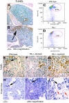
Figure 1
Figure 1
Localized necrotic cell death in Rb-null fetal liver. (A-B) TUNEL immunohistochemistry on sagittal sections of wild-type and Rb-null E13.5 fetal liver. (C-D) Representative flow cytometric analysis of annexin-V and TER119 staining on disaggregated whole E13.5 fetal liver from wild-type (C, 7.1% ± 0.6%, P < .002) and Rb-null (D, 2.8% ± 1.0%, P < .002) embryos to detect phosphotidyl serine externalization. (E-G) TUNEL staining for DNA fragmentation of wild-type (E) and Rb-null (F-G) E13.5 fetal livers (1000× magnification). The arrow in panel E identifies a phagocytic macrophage and s indicates a liver sinusoid. The dorsal region of the Rb-null fetal liver (F) shows markedly less TUNEL positivity than the ventral end of the liver that is distant from the hepatic vasculature. (H-J) Protein blue staining of sections of E13.5 wild-type fetal liver (H), more dorsal, healthy regions of the Rb-null fetal liver (I), and dying regions in the ventral end of the Rb−/− fetal liver (J) with black arrows indicating the presence of phagocytic macrophages and red arrows indicating distended, remnant nuclei. See ″Fluorescence microscopy and island counts″ for image acquisition information.
Figure 2
Figure 2
Rb-null macrophages are competent for erythroblast interactions in vivo. (A-C) Immunohistochemical staining for F4/80 on Rb-null fetal liver showing the increased numbers of F4/80+ macrophages accumulating in the distal tip of the liver (red arrow, B), and the difference in staining of F4/80 in healthy regions of the liver to the right of the red line (D) compared with dying and ischemic regions of the liver to the left of the red line (C). (D-E) F4/80 immunohistochemistry on intact Rb-null (F) and wild-type (E) fetal liver with F4/80-positive surface area determined by optical densitometry for DAB positivity. (F) Graphic representation of total fetal liver cell number, number of F4/80-positive macrophages, number of TER119-positive erythroblasts, and ratio of TER119-positive erythroblasts to F4/80-positive macrophages in wild-type and Rb-null fetal livers at E12.5 and E13.5. At E12.5, F4/80-positive macrophages represented 13.2% (± 2.7%) and 15.4% (± 0.9%) of total cell number for wild type and Rb null, respectively, and 13.4% (± 4.0%) for wild-type and 12.2% (± 0.4%) for Rb null at E13.5. (G-H) Protein blue-stained E13.5 wild-type (H) and Rb-null (I) fetal livers showing erythroblastic islands in situ (left) with a color-coded key (right) to indicate distinct cell types, including macrophages (M, green), erythroblasts (E, red), hepatocytes (H, brown), endothelial cell (en, turquoise), and liver sinusoids (S). (I-J) Electron microscopy of macrophage-erythroblast contacts in E13.5 wild-type (J) and Rb-null (K) fetal liver. Erythroblasts (Ery), macrophages (Mac), and hepatocytes (Hep) were distinguished on the basis of mitochondrial size and numbers (hepatocytes have larger, more numerous mitochondria), nuclear and cytoplasmic electron density, and smaller cell size (erythroblasts) and larger cell size, cytoplasmic projections, and inclusion bodies (macrophages). See ″Fluorescence microscopy and island counts″ for image acquisition information.
Figure 3
Figure 3
Macrophages are not required for erythroid enucleation. (A,B) Native erythroblast islands from disaggregated wild-type (left) or Rb−/− (right) E12.5 fetal liver were examined 16 hours after culture, using anti-F4/80 to stain macrophages (red) and anti-TER119 (green) to stain erythroblasts, and Hoechst 33342 for DNA, indicating no difference in island formation between wild-type and Rb-null-derived cells. (C,D) High-magnification analysis of the interaction between red cells (benzidine-stained brown cells) and macrophages (large “foamy” cells) is examined in the context of 12-day CFU-GEMM colony differentiation derived from wild-type (C) or Rb-null (D) fetal liver progenitors. We noted that Rb-null erythroblasts (D) failed to enucleate irrespective of macrophage contacts (arrow) and demonstrated increased size and abnormal chromatin structure as reported previously., (E,F) Native erythroblast islands cultured in vitro from mice with the indicated Rb genotypes and at early or later stages of embryonic development were enumerated (F). Only definitive islands in which 5 or more erythroblasts were bound were counted as islands. Numbers of erythroblasts per macrophage were also counted (E). Experiments were carried out in triplicate. Error bars are standard deviations. Significant differences were observed for the number of erythroblasts per macrophage in “late” E13.5 wild-type and Rb-null fetal liver after 4 hours (P = .001) or 16 hours (P = .001) in culture (E). Student t test. (G) Quantitation of enucleation by flow cytometric analysis of all erythroid cells (floating and attached) was carried out after 60 hours in culture, either in the presence of macrophages and erythroblast island formation or in suspension culture. The percentage of enucleated Rb-null cells was significantly reduced relative to wild type in suspension culture (P < .009) and in macrophage coculture (P < .003). (H) Purified erythroblast progenitors from E12.5 fetal livers were cultured for 48 hours in adherence with macrophages or in suspension culture, and then harvested for CFU-E assay in methylcellulose. Viable cells (250) were plated in triplicate per sample. Numbers of CFU-Es formed were compared with cultures seeded with erythroblasts recovered after only 90 minutes in culture (day 0). See ″Fluorescence microscopy and island counts″ for image acquisition information.
Figure 4
Figure 4
Up-regulation of hypoxia-inducible genes in Rb-null fetal liver without changes in macrophage-specific or PU1 target genes. Quantitative real-time PCR using wild-type and Rb-null total E13.5 fetal liver cDNA was used to validate and extend data from differential microarray analysis of gene expression. Values are the mean value obtained from 3 separate data sets for each genotype with the wild-type values normalized to 1.0 and values for Rb null samples expressed relative to wild-type. Error bars represent the standard deviation from the mean for each genotype.
Figure 5
Figure 5
Hypoxia disrupts erythropoietic islands. (A,B) The effect of hypoxia on erythropoietic island numbers and numbers of erythroblasts per macrophage was quantified as described previously (Figure 3E,F). Hypoxia had a greater effect on both island integrity and numbers of erythroblasts per macrophage than did the Rb status of the cells. Differences in numbers of erythroblasts per macrophage as a function of oxygen tension (21% vs 1% O2) were significant in the absence of Cre expression (P < .001) and in the presence of Cre expression (P < .001). The numbers of islands formed at 21% compared with 1% O2 were also significant (P < .04 for Cre− and P < .02 for Cre+). Error bars represent standard deviation from the mean value for numbers of erythroblasts per island (A) and numbers of islands (B). (C-H) The effect of hypoxia on erythroblast island integrity was visualized by staining of adherent (C-F) and floating (G,H) cells from purified island cultures exposed to 21% oxygen for 36 hours (C,D) or to 21% oxygen for 18 hours and subsequently to 1.0% oxygen for 18 hours (E-H). Significant differences were observed for the number of erythroblasts per macrophage in E13.5 wild-type and Rb-null fetal liver after 4 hours (P = .001) or 16 hours (P = .001) in culture at 21% oxygen and between Rb-positive and Rb-deficient cultures derived from Rbflox mice when cultured at 1% oxygen versus 21% oxygen for 12 hours (P = .001 for Cre+; P = .001 for Cre−) but not between wild type and Rb null earlier in gestation or possessing a wild-type placenta (Rbflox;Cre+ vs Cre−). See ″Fluorescence microscopy and island counts″ for image acquisition information. (B) Island numbers were reduced in E13.5 Rb-null fetal liver compared with wild type (P = .01) and in conditionally targeted fetal livers following 16 hours at 1% oxygen when compared with normal tissue culture (P = .02 and P = .04, respectively) Notably, placental rescue increased Rb-deficient island number (P = .03; Student t test).
Figure 6
Figure 6
Model: a putative role for macrophages in the homeostatic response to hypoxia, based on in vitro analyses. In response to hypoxia, we propose that erythroblasts lose their tight adherence to central macrophages of erythroblastic islands, possibly promoting unrestrained maturation of erythroid progenitors, and increasing the number of red cells getting into the circulation. This may also promote the migration of immature erythroid progenitors to organs such as the spleen where their expansion is enhanced under stress conditions. In the Rb-null embryo, severe hypoxia and fetal liver necrosis disrupt erythropoietic islands concomitant with, but independent of, red cell enucleation defects and embryonic lethality.

Similar articles

Cited by

References

    1. Bessis M. L'ilot erythroblastique: unite functionelle de la moelle osseuse. Rev Hematol. 1958;13:8–11. - PubMed
    1. Sadahira Y, Mori M. Role of the macrophage in erythropoiesis. Pathol Intl. 1999;49:841–848. - PubMed
    1. Chasis JA. Erythroblastic islands: specialized microenvironmental niches for erythropoiesis. Curr Opin Hematol. 2006;13:137–141. - PubMed
    1. Sasaki K, Iwatsuki H, Suda M, Itano C. Scavenger macrophages and central macrophages of erythroblastic islands in liver hematopoiesis of the fetal and early post-natal mouse: a semithin light- and electron-microscopic study. Acta Anat. 1993;147:75–82. - PubMed
    1. Hanspal M, Hanspal JS. The association of erythroblasts with macrophages promotes erythroid proliferation and maturation: a 30 kD heparin-binding protein is involved in this contact. Blood. 1994;84:3494–3504. - PubMed

Publication types