. 2007 Oct;116(5):471-86.
doi: 10.1007/s00412-007-0111-y. Epub 2007 Jun 9.

Interactions between Mei4, Rec114, and other proteins required for meiotic DNA double-strand break formation in Saccharomyces cerevisiae

Affiliations

Interactions between Mei4, Rec114, and other proteins required for meiotic DNA double-strand break formation in Saccharomyces cerevisiae

Shohreh Maleki et al. Chromosoma. 2007 Oct.

Abstract

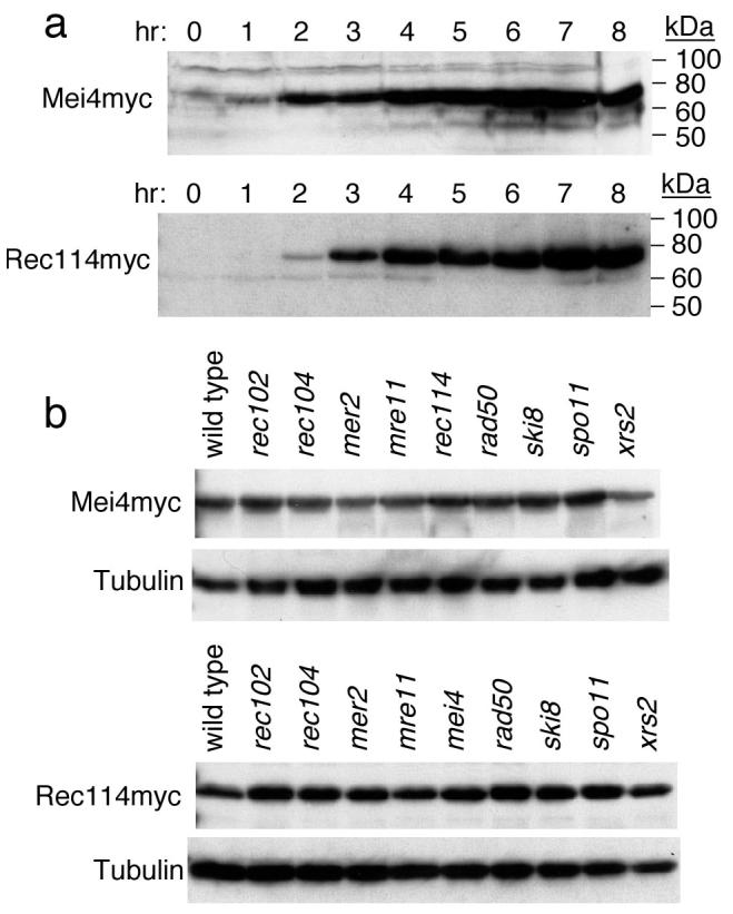
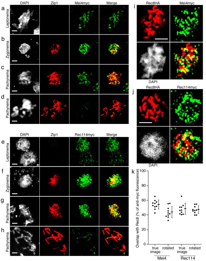
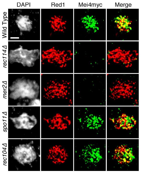
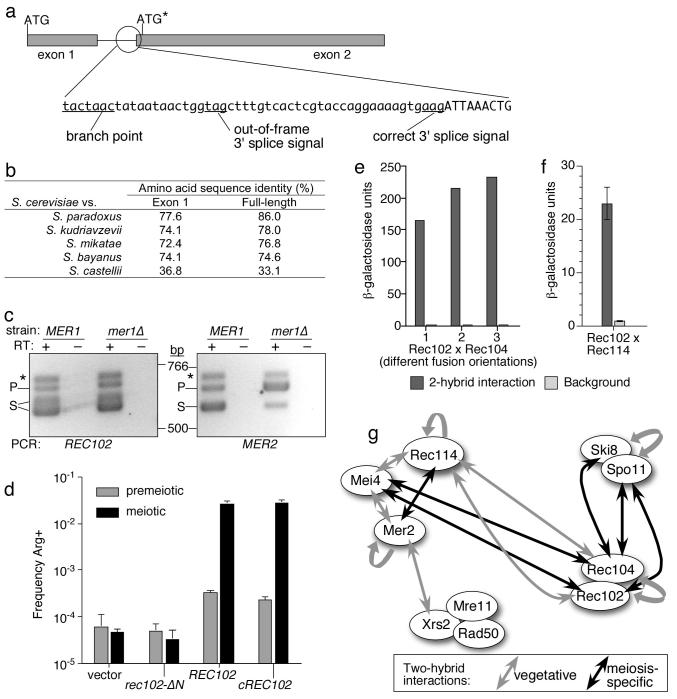
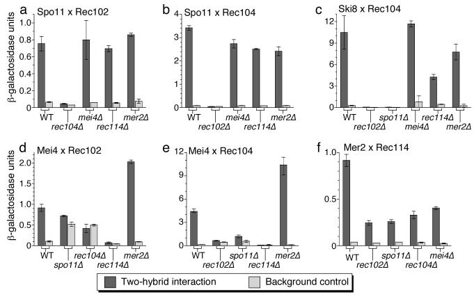
In most sexually reproducing organisms, meiotic recombination is initiated by DNA double-strand breaks (DSBs) formed by the Spo11 protein. In budding yeast, nine other proteins are also required for DSB formation, but the roles of these proteins and the interactions among them are poorly understood. We report further studies of the behaviors of these proteins. Consistent with other studies, we find that Mei4 and Rec114 bind to chromosomes from leptonema through early pachynema. Both proteins showed only limited colocalization with the meiotic cohesin subunit Rec8, suggesting that Mei4 and Rec114 associated preferentially with chromatin loops. Rec114 localization was independent of other DSB factors, but Mei4 localization was strongly dependent on Rec114 and Mer2. Systematic deletion analysis identified protein regions important for a previously described two-hybrid interaction between Mei4 and Rec114. We also report functional characterization of a previously misannotated 5' coding exon of REC102. Sequences encoded in this exon are essential for DSB formation and for Rec102 interaction with Rec104, Spo11, Rec114, and Mei4. Finally, we also examined genetic requirements for a set of previously described two-hybrid interactions that can be detected only when the reporter strain is induced to enter meiosis. This analysis reveals new functional dependencies for interactions among the DSB proteins. Taken together, these studies support the view that Mei4, Rec114, and Mer2 make up a functional subgroup that is distinct from other subgroups of the DSB proteins: Spo11-Ski8, Rec102-Rec104, and Mre11-Rad50-Xrs2. These studies also suggest that an essential function of Rec102 and Rec104 is to connect Mei4 and Rec114 to Spo11.


Figures

Fig. 1
Fig. 1
Epitope-tagged Mei4 and Rec114 proteins. (a) Western blot analysis of time courses of Mei4myc and Rec114myc expression. Migration positions of protein size standards are shown. (b) Steady-state levels of Mei4myc and Rec114myc in DSB-defective mutants. Whole-cell extracts at 4 hr in meiosis were analyzed by anti-myc western blot. Blots probed with anti-tubulin antibody served as loading controls.
Fig. 2
Fig. 2
Temporal and spatial patterns of Mei4 and Rec114 association with chromosomes. (a–d) Localization of Mei4. (e–h) Localization of Rec114. Nuclear spreads from meiotic cultures of MEI4myc or REC114myc strains were stained with anti-Zip1 (red) and anti-myc (green) antibodies. Equivalent exposures of representative nuclei are shown, staged according to the extent of SC formation. (i–k) Localization of Mei4 or Rec114 with respect to chromosome axes. Nuclear spreads of MEI4myc REC8HA (i) or REC114myc REC8HA (j) strains were stained with anti-HA (red) and anti-myc (green) antibodies. Equivalent exposures of representative late zygotene nuclei are shown. (k) Quantification of the extent of overlap of Mei4myc or Rec114myc immunofluorescence with Rec8HA. Overlap was also assessed for the same nuclei with the green fluorescence channel rotated 180° in order to estimate random colocalization. Error bars are mean ± s.d. Scale bars, 2 μm.
Fig. 3
Fig. 3
Genetic requirements for stable chromosomal association of Mei4myc. Nuclear spreads were prepared from meiotic cultures of a MEI4myc strain (wild type) and MEI4myc strains carrying the indicated DSB gene mutations. Zip1 staining could not be used to stage the nuclei because DSB-defective mutants are defective for SC formation. Therefore, spreads were double-stained for Mei4myc (green) and a different chromosome structure protein, Red1 (red). Exposures equivalent to the ones in Fig. 2 are shown. Scale bar, 2 μm.
Fig. 4
Fig. 4
Deletion analysis of the interaction between Rec114 and Mei4. (a) Serial deletions were constructed containing the indicated amino acid residues of Mei4 fused to the C terminus of LexA. For comparison, the schematic of S. cerevisiae Mei4 showing conservation with homologs from other yeasts is reproduced from Fig. S1. Each construct was assayed in vegetatively growing reporter cells for two-hybrid interaction with full-length Rec114. Units of β-galactosidase expressed when the LexA-Mei4 construct was paired with Gal4AD-Rec114 and when paired with empty Gal4AD vector (“Control”) are shown (single measurement for each). (b) Western blot analysis of steady-state expression levels for full-length LexA-Mei4 and interaction-defective N-terminal deletions. Bands corresponding to LexA-Mei4 fusion proteins are marked with open circles; asterisks indicate cross-reacting bands. Extract from equivalent numbers of cells was loaded in each lane. (c) As for panel (a), except serial deletions of Rec114 fused to the C terminus of Gal4AD were assayed for interaction with full-length Mei4. (d) Western blots probed with anti-HA antibody to detect full-length and interaction-defective Gal4AD-Rec114 fusion proteins, with or without co-expression of LexA-Mei4 as indicated (LexA-Mei4 was not detected in this blot). Different amounts of extract were loaded because the smaller fusion proteins accumulated to higher steady-state levels than the full-length fusion. Gal4AD fusions (or Gal4AD alone) are marked with open circles; the asterisk marks a set of nonspecific cross-reacting bands apparent when larger amounts of extract were loaded. The reason the fusion proteins migrate as doublets or smears is unknown, but it may reflect proteolysis, translational read-through of the stop codon, and/or post-translational modification.
Fig. 5
Fig. 5
REC102 genomic structure and requirement for the 5′ exon. (a) Schematic of REC102 genomic locus. Exons are denoted by gray boxes and the intron by a horizontal line. Positions of the correct ATG and the previously annotated start codon (asterisk) are indicated. The circled area highlights the genomic sequence around the 3′ end of the intron (lower case, intron sequence; upper case, coding sequence). The two alternative 3′ splice signals are indicated. (b) Sequence conservation of Rec102 homologs. The amino acid sequence of S. cerevisiae Rec102 was compared to that of homologs from the indicated Saccharomyces species, both for the full-length protein and for just the sequence encoded in exon 1. (c) RT-PCR analysis of REC102. Total RNA was isolated from wild-type and mer1Δ strains six hr after transfer to sporulation medium. REC102 (left panel) and MER2 (right panel) were amplified by RT-PCR with (+) or without (−) reverse transcriptase, as indicated. Negative images of an ethidium-stained gel are shown. PCR products from unspliced precursor RNA (P) and spliced mRNA (S) are shown. The bands marked by asterisks are template-switching artifacts generated by the reverse transcriptase (data not shown). (d) Exon 1 is required for REC102 function. Return-to-growth assays were conducted in a rec102Δ strain carrying the indicated REC102 sequences under the control of a constitutive promoter on a 2μ plasmid. Values are the mean ± sd for three independent cultures. Premeiotic recombinant frequencies are often lower for DSB-deficient strains because some Arg+ recombinants arise from cells that prematurely entered meiosis during the premeiotic growth regimen. (e) Two-hybrid interactions of full-length Rec102 with Rec104 were assayed in vegetative cultures in various orientations: 1, LexA-Rec102 × Gal4AD-Rec104; 2, LexA-Rec102 × Rec104-Gal4AD; 3, Rec104-LexA × Gal4AD-Rec102 (single measurement of each). Background controls contained the LexA fusion protein plus Gal4AD alone. For comparison, interaction of LexA-Rec102ΔN and Gal4AD-Rec104 yielded less than 0.8 units of β-galactosidase (Kee et al. 2004). (f) Two-hybrid interaction of full-length LexA-Rec102 with Gal4AD-Rec114 was assayed in vegetative cultures. Error bars are mean ± range for two cultures. (g) Two-hybrid interaction network, incorporating new Rec102 interactions (adapted from Arora et al. 2004). Gray arrows are interactions observed in a vegetative reporter strain. Black arrows are interactions observed only in a meiotic reporter strain. For clarity, the figure omits interactions among Rad50, Mre11 and Xrs2 and also omits several interactions that gave relatively weak LacZ signals (less than 10-fold over background) that have not been evaluated by independent methods (see Arora et al. 2004 for further details).
Fig. 6
Fig. 6
Genetic requirements for meiosis-specific two-hybrid interactions. Each reporter strain carried a deletion of one of the meiosis-specific DSB genes, as indicated. (“WT”, wild type for the DSB genes. Note that all strains also carry the ndt80 mutation to block meiosis in prophase I.) Dark gray bars are two-hybrid signals for LexA and Gal4AD fusions to the indicated DSB proteins; light gray bars are controls carrying the LexA fusion with an empty Gal4AD vector. Values are the mean ± range for 2–3 independent measurements. Variation was low enough that some error bars are not visible. Fusion orientations were as follows: (a) Spo11-LexA × Gal4AD-Rec102; (b) Spo11-LexA × Rec104-Gal4AD; (c) LexA-Ski8 × Rec104-Gal4AD; (d) LexA-Mei4 × Gal4AD-Rec102; (e) LexA-Mei4 × Rec104-Gal4AD; (f) LexA-Mer2 × Gal4AD-Rec114.

Similar articles

Cited by

References

    1. Alani E, Padmore R, Kleckner N. Analysis of wild-type and rad50 mutants of yeast suggests an intimate relationship between meiotic chromosome synapsis and recombination. Cell. 1990;61:419–436. - PubMed
    1. Araki Y, Takahashi S, Kobayashi T, Kajiho H, Hoshino S, Katada T. Ski7p G protein interacts with the exosome and the Ski complex for 3'-to-5' mRNA decay in yeast. EMBO J. 2001;20:4684–4693. - PMC - PubMed
    1. Arora C, Kee K, Maleki S, Keeney S. Antiviral protein Ski8 is a direct partner of Spo11 in meiotic DNA break formation, independent of its cytoplasmic role in RNA metabolism. Mol Cell. 2004;13:549–559. - PubMed
    1. Assenmacher N, Hopfner KP. MRE11/RAD50/NBS1: complex activities. Chromosoma. 2004;113:157–166. - PubMed
    1. Blat Y, Protacio RU, Hunter N, Kleckner N. Physical and functional interactions among basic chromosome organizational features govern early steps of meiotic chiasma formation. Cell. 2002;111:791–802. - PubMed

Publication types