

Review
doi: 10.1002/jcp.21172.
Liver regeneration
Affiliations
- PMID: 17559071
- PMCID: PMC2701258
- DOI: 10.1002/jcp.21172
Item in Clipboard
Review
Liver regeneration
J Cell Physiol.
2007 Nov.
Abstract
Liver regeneration after partial hepatectomy is a very complex and well-orchestrated phenomenon. It is carried out by the participation of all mature liver cell types. The process is associated with signaling cascades involving growth factors, cytokines, matrix remodeling, and several feedbacks of stimulation and inhibition of growth related signals. Liver manages to restore any lost mass and adjust its size to that of the organism, while at the same time providing full support for body homeostasis during the entire regenerative process. In situations when hepatocytes or biliary cells are blocked from regeneration, these cell types can function as facultative stem cells for each other.
Figures
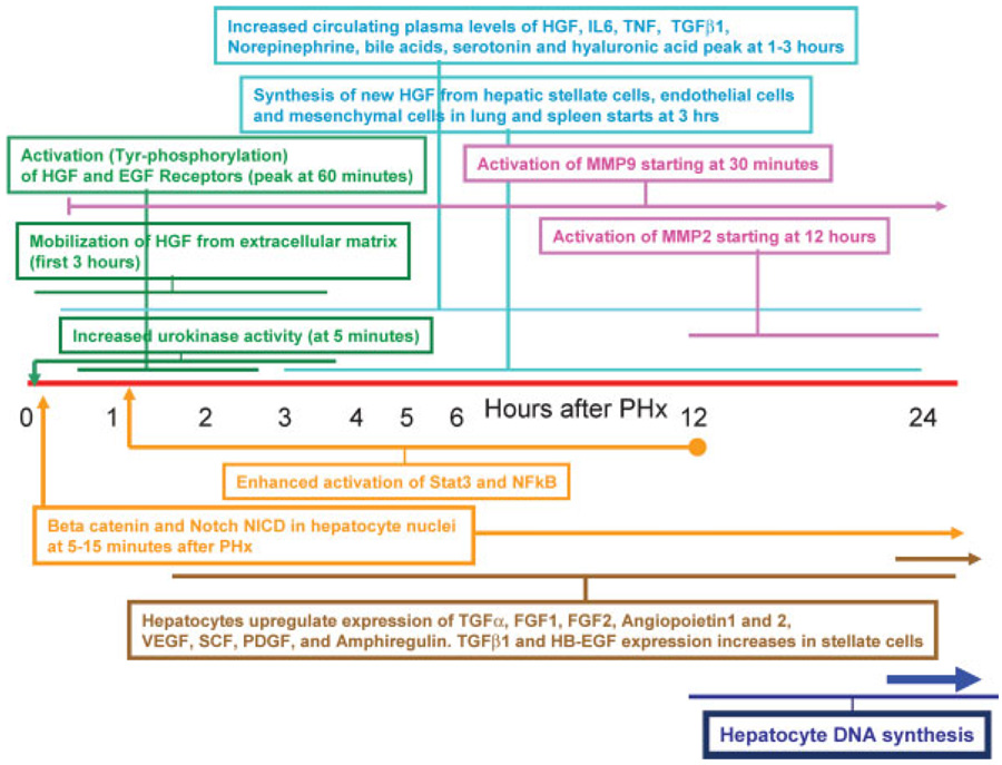
Chronology of key events occurring at the early stages of liver regeneration after partial hepatectomy. Events within similarly colored boxes belong in the same category (e.g., green: growth factor related events; blue: plasma related changes, etc.). The associated horizontal lines for each box delineate the beginning and the duration of each signal.
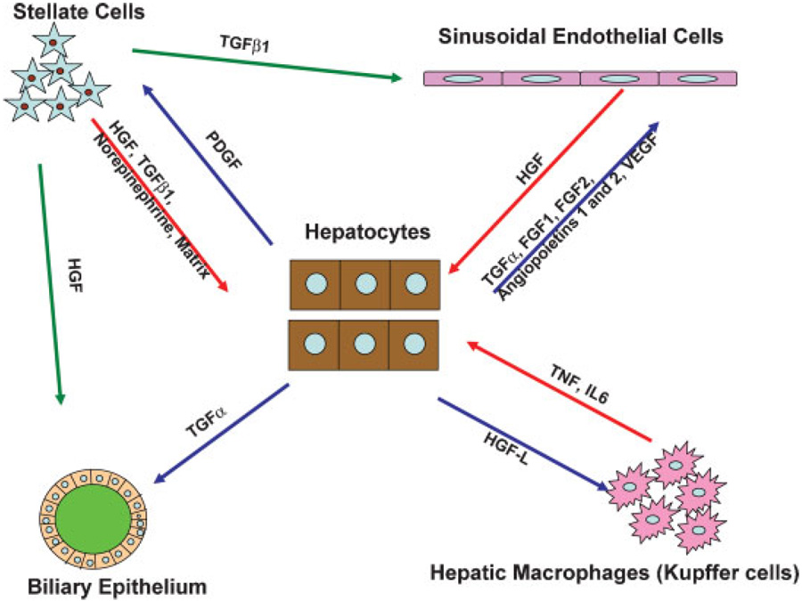
Signaling interactions between different hepatic cell types during liver regeneration.
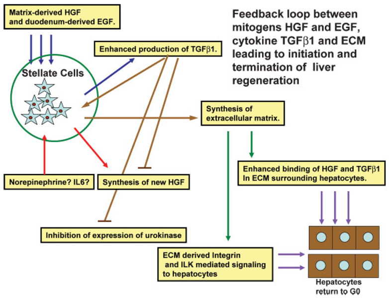
Schematic of a feedback loop between growth factors, TGFβ1, and extracellular matrix, controlling early and late stages of regeneration. Mitogens (HGF and EGF) upregulate expression of TGFβ1 by stellate cells. The latter stimulates synthesis of new extracellular matrix, while eventually blocking synthesis of new HGF and expression of urokinase. The newly synthesized extracellular matrix supports binding of single chain HGF and TGFβ1 around hepatocytes and restoration of quiescence (G0 phase). Arrows of the same color denote similar origin of the input and output of the same signaling process.
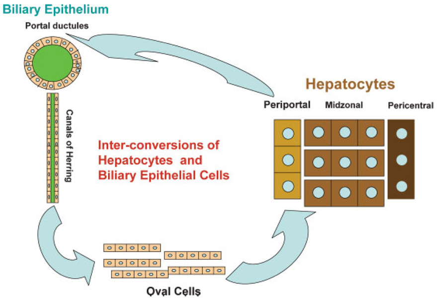
Cells from the biliary compartment (portal ductules and canals of Herring) transform into oval cells and these become hepatocytes when proliferation of hepatocytes is inhibited during regeneration. Periportal hepatocytes can also convert to biliary cells when there is injury to biliary cells but their capacity for self-repair is inhibited. Hepatocytes and biliary cells are facultative stem cells for each other.
References
-
- Aaronson SA, Rubin JS, Finch PW, Wong J, Marchese C, Falco J, Taylor WG, Kraus MH. Growth factor-regulated pathways in epithelial cell proliferation. Am Rev Respir Dis. 1990;142:S7–S10. - PubMed
-
- Akerman P, Cote P, Yang SQ, McClain C, Nelson S, Bagby GJ, Diehl AM. Antibodies to tumor necrosis factor-alpha inhibit liver regeneration after partial hepatectomy. Am J Physiol. 1992;263:G579–G585. - PubMed
-
- Alison MR, Poulsom R, Forbes SJ. Update on hepatic stem cells. Liver. 2001;21:367–373. - PubMed
-
- Appasamy R, Tanabe M, Murase N, Zarnegar R, Venkataramanan R, Van Thiel DH, Michalopoulos GK. Hepatocyte growth factor, blood clearance, organ uptake, and biliary excretion in normal and partially hepatectomized rats. Lab Invest. 1993;68:270–276. - PubMed
-
- Argast GM, Campbell JS, Brooling JT, Fausto N. Epidermal growth factor receptor transactivation mediates tumor necrosis factor-induced hepatocyte replication. J Biol Chem. 2004;279:34530–34536. - PubMed
Publication types
MeSH terms
Substances
Grants and funding
LinkOut - more resources
Full Text Sources
Other Literature Sources
Medical
