. 2007 Jul 15;110(2):420-31.
doi: 10.1002/cncr.22781.

Generation and characterization of an ascitogenic mesothelin-expressing tumor model

Affiliations

Generation and characterization of an ascitogenic mesothelin-expressing tumor model

Wen-Fang Cheng et al. Cancer. .

Abstract

Background: Intraperitoneal tumors expressing high amounts of mesothelin such as malignant mesothelioma and ovarian cancers tend to develop ascites and result in significant morbidity and mortality in the patient. A suitable preclinical intraperitoneal model will assist in the illustration of the mechanisms of molecular oncogenesis and facilitate in addressing issues related to early screening, diagnosis, and therapy for intraperitoneal tumors.

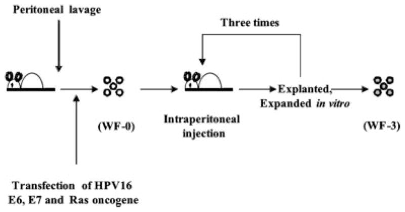
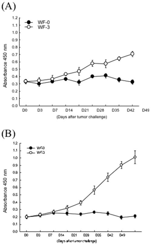
Methods: In the current study, an ascitogenic malignant tumor model (WF-3) was created. The mobility and proliferation of WF-3 and its precursor cells, WF-0, were characterized using transwell and MTT (3-(4,5-dimethylthiazol-2-yl)-2,5-diphenyltetrazolium bromide) assays. In addition, the in vivo tumorgenicity of WF-3 and WF-0 was determined using intraperitoneal injection of the tumor cells. Microarray analysis was performed using WF-3 and WF-0. Northern blot analysis was used to characterize the expression of the mesothelin gene in WF-3 and WF-0. Furthermore, the mesothelin levels in serum and ascites were used to correlate with tumor load of WF-3 in tumor challenged mice.

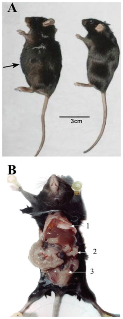
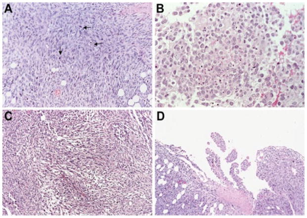
Results: The WF-3 tumor cells demonstrated relatively high proliferation and migration rates compared with the parental cell line, WF-0. The tumors from the WF-3 but not WF-0 were capable of forming ascites and peritoneal-based tumors after tumor challenge. The WF-3 tumor model was also capable of implanting into multiple organs including the diaphragm, intestines, and peritoneal wall. Furthermore, the WF-3 tumor expressed high levels of mesothelin, which is commonly observed in the majority of ovarian cancers, pancreatic cancer, and malignant mesothelioma. In addition, the authors found that the serum and ascites mesothelin levels correlated with tumor loads in tumor-challenged mice.

Conclusions: The data indicate that the WF-3 murine tumor model may potentially serve as a good model for understanding the molecular oncogenesis of peritoneal tumors. In addition, the preclinical model may potentially be useful for the development of diagnostic and therapeutic methods against intraperitoneal cancers.


Figures

FIGURE 1
FIGURE 1
Schematic diagram showing the in vivo selection of WF-0, WF-1, WF-2, and WF-3 cell lines. The peritoneal cells of the C57BL/6 mice were collected, transduced with retrovirus encoding human papillomavirus type 16 (HPV-16) E6 and E7 genes, followed by transfection with DNA encoding human c-Ha-ras gene, and named WF cells. The WF cells were injected intraperitoneally into athymic mice repeatedly. Eventually, athymic mice developed ascites. The tumor cells from ascites in athymic mice previously challenged with WF cells were isolated, expanded in vitro, and named WF-0. When WF-0 cells were injected into C57BL/6 mice, <10% of WF-0 tumor-challenged C57BL/6 mice developed ascites. Tumor cells from the ascites of C57BL/6 mice were isolated and expanded in vitro. These expanded cell lines were called WF-1. The mice were further challenged intraperitoneally with WF-1. The outgrown ascites in mice challenged with WF-1 were further cultured in vitro. These expanded cell lines were called WF-2. The mice were challenged intraperitoneally with WF-2. Cells harvested from the ascites of mice challenged with WF-2 were further cultured in vitro. These expanded cell lines were called WF-3.
FIGURE 2
FIGURE 2
In vitro tumor cell migration assays. The migration of tumor cells was assessed by counting the number of cells that migrated through transwell. The cells that migrated through the membranes into the lower wells were assessed by hematoxylin uptake and the number of cells was counted, or by the MTT (3-(4,5-dimethylthiazol-2-yl)-2,5-diphenyltetrazolium bromide) assay. (A) Representative figures of cell migration in WF-0 and WF-3 cells. (B) Bar graph depicting total number of tumor cells that migrated through transwell. Note that the cell numbers that migrated through the membranes in the WF-3 group were significantly higher than those in the WF-0 group (P < .05, 1-way analysis of variance [ANOVA]). (C) Bar graph depicting absorbance at 570 nanometers (nm) using enzyme-linked immunoadsorbent assay (ELISA) in WF-0 or WF-3 tumor cells treated with MTT solution. Note that the absorbance at 570 nm of the WF-3 group were also significantly higher than those of the WF-0 group (P < .05, 1-way ANOVA).
FIGURE 3
FIGURE 3
In vitro measurement of cell proliferation and in vivo tumor growth experiments. We characterized the proliferative rates of WF-0 and WF-3 cell lines in vitro. The WF-0 and WF-3 cells were seeded with the same number of tumor cells. The tumor cells were collected at different time points and characterized by MTT (3-(4,5-dimethylthiazol-2-yl)-2,5-diphenyltetrazolium bromide) assays (A) or direct cell counting under the microscope (B). Note that the absorption at OD 570 nanometers (nm) of the WF-3 cells was significantly higher than that of the WF-0 cells (P < .05, 1-way analysis of variance [ANOVA]); the total numbers of the WF-3 cells were significantly higher than those of the WF-0 cells (P < .05, 1-way ANOVA). (C) In vivo tumor growth experiments. C57BL/6 mice were challenged with WF-0 (closed circle) or WF-3 (open circle) subcutaneously. Tumor growth was measured twice a week. Note that no subcutaneous tumor growth was identified in mice challenged with WF-0. In contrast, all of the mice challenged with WF-3 developed tumor growth. SD indicates standard deviation.
FIGURE 4
FIGURE 4
Ascitogenic features of mice challenged with WF-3 cells intraperitoneally. C57BL/6 mice were challenged with 5 × 10⁴/mouse of WF-3 tumor cells intraperitoneally. The tumor-challenged mice were sacrificed 12 weeks after tumor challenge. (A) Representative figures of tumor-challenged mice showing ascites formation. The mice challenged with WF-3 cells were found to have a distended abdomen (left mouse). In contrast, the control mouse without tumor challenge revealed a scaphoid abdomen (right mouse). (B) Representative figures showing the distribution of WF-3 tumors in the peritoneal cavity. Note: multiple, friable, and grayish-white tumor nodules of various sizes were found in the peritoneal cavity. The tumor implants were identified in multiple abdominal organs, including diaphragm (arrow 1), peritoneal wall (arrow 2), and intestine (arrow 3).
FIGURE 5
FIGURE 5
Morphologic features of WF-3 tumor cells. WF-3 tumor cells were challenged intraperitoneally into C57BL/6 mice at a dose of 5 × 10⁴/mouse. The mice were sacrificed 12 weeks after the tumor challenge. Tissue sections derived from the tumors were prepared by hematoxylin and eosin staining. The morphology as well as the invasiveness of the tumor was characterized by histology. (A) The tumors were characterized by sheets of bizarre anaplastic tumor cells with marked pleomorphism, a high nuclear:cytoplasmic ratio, and hyperchromatism with numerous mitotic figures (arrows). (B) Some areas of the WF-3 tumors showed a carcinomatous component. (C) Some areas of the WF-3 tumor showed a sarcomatous component. (D) Focal areas of the WF-3 tumor also demonstrated papillary configuration.
FIGURE 6
FIGURE 6
In vivo tumor growth kinetics of WF-0 and WF-3 cells. C57BL/6 mice were challenged with different doses of WF-0 or WF-3 tumor cells (1 × 10⁴, 5 × 10⁴, or 1 × 10⁵/mouse) intraperitoneally. Tumor growth was monitored twice a week. In addition, survival of mice was characterized by Kaplan-Meier survival analysis. (A) In vivo tumor growth experiments in mice challenged with WF-0 cells. (B) Survival analysis in mice challenged with WF-0 cells. (C) In vivo tumor growth experiments in mice challenged with WF-3 cells. (D) Survival analysis in mice challenged with WF-3 cells. Note that all of the mice challenged with WF-3 tumor cells developed tumors and ascites within 25 days after challenging with 5 × 10⁴ or 1 × 10⁵/mouse WF-3 tumor cells. 80% of mice challenged with WF-3 at a dose of 1 × 10⁴ developed a tumor 49 days after tumor challenge. In contrast, none of the mice challenged with WF-0 developed tumor growth. Furthermore, all of the mice challenged with 5 × 10⁴ or 1 × 10⁵/mouse of WF-3 tumor cells died within 60 days after tumor challenge. 80% of mice challenged with 1 × 10⁴/mouse of WF-3 tumor cells died within 90 days after tumor challenge. In contrast, none of the mice challenged with WF-0 died 90 days after tumor challenge.
FIGURE 7
FIGURE 7
Northern blot analysis to characterize mesothelin expression in WF-0 and WF-3 cells. Total cellular RNA from cultured WF-0 and WF-3 tumor cells were isolated. A 20-μg portion of total RNA was resolved on a 1.5% formaldehyde-agarose gel (B) and blotted onto a nylon membrane (A). The hybridization was then performed with 32P-labeled mouse mesothelin DNA fragment probe generated by the random-priming method. Note: Equal amounts of RNA from WF-0 and WF-3 tumor cells were loaded onto the wells as indicated by the same amounts of 28S and 18S RNA (indicated by arrows, B). The expression level of mesothelin in WF-3 cells is greater than that of the WF-0 cells (indicated by arrow, A).
FIGURE 8
FIGURE 8
Titers of mesothelin in sera and ascites of mice challenged with WF-0 and WF-3 cells. To evaluate the levels of mesothelin in the sera or ascites of mice challenged with WF-0 or WF-3 cells, we performed enzyme-linked immunoadsorbent assay (ELISA). (A) ELISA assay characterizing serum mesothelin levels in WF-0 (closed circle) or WF-3 (open circle) tumor challenged mice. (B) ELISA assay characterizing ascites mesothelin levels in WF-0 (closed circle) or WF-3 (open circle) tumor-challenged mice. Note that both the serum and ascites mesothelin levels in WF-3 tumor-challenged mice were significantly higher than those of WF-0 tumor-challenged mice at 14 days after tumor challenge (P < .05, 1-way analysis of variance [ANOVA]). nm indicates nanometers.
FIGURE 9
FIGURE 9
Characterization of body weights and mesothelin level in serum and ascites after WF-3 tumor cell challenge. Mice were challenged with WF-3 tumor or 1× phosphate-buffered saline (PBS). The body weight of the challenged mice was followed over time. Furthermore, the serum and ascites mesothelin levels of the WF-3 tumor-challenged mice were characterized by enzyme-linked immunoadsorbent assay (ELISA). (A) Body weight measurements after WF-3 tumor (open circle) or PBS (closed circle) challenge. Note that the WF-3 tumor-challenged mice gained significant weight compared with PBS-challenged mice starting 15 days after challenge. (B) Correlations between body weights and mesothelin levels in serum (closed circle) or ascites (open circle) of WF-3-injected mice. Note that the body weights appear to correlate with the serum or ascitic mesothelin levels in WF-3 tumor-challenged mice more obviously when the mice weight is >25 g. SD indicates standard deviation; nm, nanometers.

References

    1. Greenlee RT, Murray T, Bolden S, Wingo PA. Cancer statistics, 2000. CA Cancer J Clin. 2000;50:7–33. - PubMed
    1. Schink JC. Current initial therapy of stage III and IV ovarian cancer: challenges for managed care. Semin Oncol. 1999;26:2–7. - PubMed
    1. Zellos LS, Sugarbaker DJ. Multimodality treatment of diffuse malignant pleural mesothelioma. Semin Oncol. 2002;29:41–50. - PubMed
    1. Nowak AK, Lake RA, Kindler HL, Robinson BW. New approaches for mesothelioma: biologics, vaccines, gene therapy, and other novel agents. Semin Oncol. 2002;29:82–96. - PubMed
    1. Liu J, Yang G, Thompson-Lanza JA, et al. A genetically defined model for human ovarian cancer. Cancer Res. 2004;64:1655–1663. - PubMed

Publication types