

Antenatal dexamethasone treatment leads to changes in gene expression in a murine late placenta
- PMID: 17559929
- PMCID: PMC2040329
- DOI: 10.1016/j.placenta.2007.04.002
Antenatal dexamethasone treatment leads to changes in gene expression in a murine late placenta
Abstract
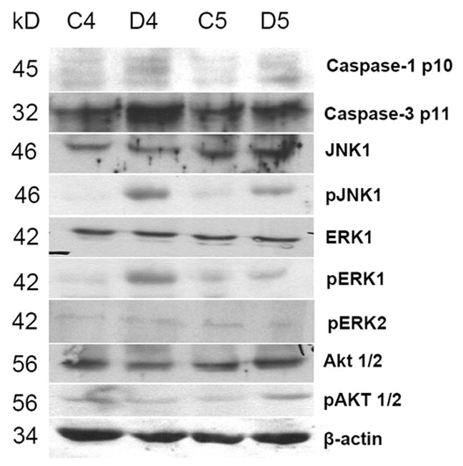
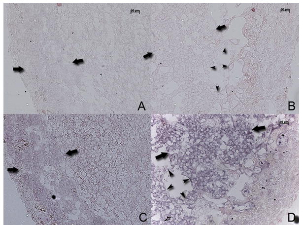
Antenatal steroids like dexamethasone (DEX) are used to augment fetal lung maturity and there is a major concern that they impair fetal growth. If delivery is delayed after using antenatal DEX, placental function and hence fetal growth may be compromised even further. To investigate the effects of DEX on placental function, we treated 9 pregnant C57/BL6 mice with DEX and 9 pregnant mice were injected with saline to serve as controls. Placental gene expression was studied using microarrays in 3 pairs and other 6 pairs were used to confirm microarray results by semi-quantitative RT-PCR, real-time PCR, in situ hybridization, western blot analysis and Oligo ApopTaq assay. DEX-treated placentas were hydropic, friable, pale, and weighed less (80.0+/-15.1mg compared to 85.6.8+/-7.6mg, p=0.05) (n=62 placentas). Fetal weight was significantly reduced after DEX use (940+/-32mg compared to 1162+/-79mg, p=0.001) (n=62 fetuses). There was >99% similarity within and between the three gene chip data sets. DEX led to down-regulation of 1212 genes and up-regulation of 1382 genes. RT-PCR studies showed that DEX caused a decrease in expression of genes involved in cell division such as cyclins A2, B1, D2, cdk 2, cdk 4 and M-phase protein kinase along with growth-promoting genes such as EGF-R, BMP4 and IGFBP3. Oligo ApopTaq assay and western blot studies showed that DEX-treatment increased apoptosis of trophoblast cells. DEX-treatment led to up-regulation of aquaporin 5 and tryptophan hydroxylase genes as confirmed by real-time PCR, and in situ hybridization studies. Thus antenatal DEX treatment led to a reduction in placental and fetal weight, and this effect was associated with a decreased expression of several growth-promoting genes and increased apoptosis of trophoblast cells.
Figures


References
-
- Crowley P. Prophylactic corticosteroids for preterm birth. Cochrane Database Syst Rev. 2000:CD000065. - PubMed
-
- Committee on Obstetric P. ACOG committee opnion: antenatal corticosteroid therapy for fetal maturation. Obstet Gynecol. 2002;99:871–873. - PubMed
-
- Moise AA, Wearden ME, Kozinetz CA, Gest AL, Welty SE, Hansen TN. Antenatal steroids are associated with less need for blood pressure support in extremely premature infants. Pediatrics. 1995;95:845–850. - PubMed
-
- Doyle LW, Ford GW, Rickards AL, Kelly EA, Davis NM, Callanan C, Olinsky A. Antenatal corticosteroids and outcome at 14 years of age in children with birth weight less than 1501 grams. Pediatrics. 2000;106:E2. - PubMed
-
- Schaap AH, Wolf H, Bruinse HW, Smolders-De Haas H, Van Ertbruggen I, Treffers PE. Effects of antenatal corticosteroid administration on mortality and long-term morbidity in early preterm, growth-restricted infants. Obstet Gynecol. 2001;97:954–960. - PubMed
Publication types
MeSH terms
Substances
Grants and funding
LinkOut - more resources
Full Text Sources
Molecular Biology Databases
Research Materials
Miscellaneous
