Review
. 2007 Aug;19(4):262-71.
doi: 10.1016/j.smim.2007.04.006. Epub 2007 Jun 7.

TCR recognition of peptide/MHC class II complexes and superantigens

Affiliations
Review

TCR recognition of peptide/MHC class II complexes and superantigens

Eric J Sundberg et al. Semin Immunol. 2007 Aug.

Abstract

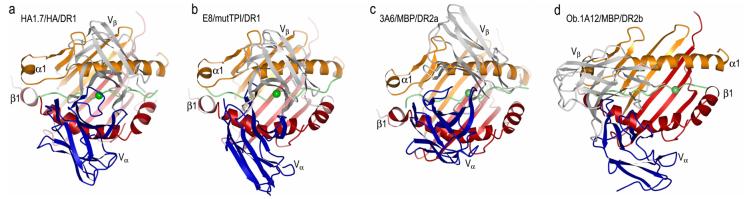
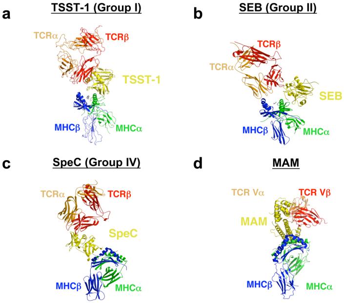
Major histocompatibility complex (MHC) class II molecules display peptides to the T cell receptor (TCR). The ability of the TCR to discriminate foreign from self-peptides presented by MHC molecules is a requirement of an effective adaptive immune response. Dysregulation of this molecular recognition event often leads to a disease state. Recently, a number of structural studies have provided significant insight into several such dysregulated interactions between peptide/MHC complexes and TCR molecules. These include TCR recognition of self-peptides, which results in autoimmune reactions, and of mutant self-peptides, common in the immunosurveillance of tumors, as well as the engagement of TCRs by superantigens, a family of bacterial toxins responsible for toxic shock syndrome.


Figures

Fig. 1
Fig. 1
Structures of human TCR/peptide/MHC class II complexes. (a) Ribbon diagram showing a top view of the anti-microbial HA1.7/HA/DR1 complex (PDB accession code 1FYT). TCR α-chain is blue and β-chain is gray; MHC α-chain is gold and β-chain is red; peptide is green. (b) The anti-tumor E8/mutTPI/DR1 complex (2IAM). (c) The autoimmune 3A6/MBP/DR2a complex (1ZGL). (d) The autoimmune Ob.1A12/MBP/DR2b complex (1YMM). In a–d, the central P5 residue of the peptide is represented as a green sphere.
Fig. 2
Fig. 2
Position of TCR CDR3 loops over foreign, self, or mutant self peptide antigens in human TCR/peptide/MHC class II complexes. Color codes for TCR and MHC molecules are the same as Fig. 1. (a) In the HA1.7/HA/DR1 complex, CDR3α and CDR3β are positioned above the central P5 residue (arrow) of the influenza HA peptide. The peptide is drawn in ball-and-stick representation with carbon atoms in green, nitrogen atoms in blue, and oxygen atoms in red. (b) In the E8/mutTPI/DR1 complex, the CDR3 loops are centered over the P3 residue (arrow) of the mutant TPI self peptide, while maintaining contacts with P5. In the 3A6/MBP/DR2a (c) and Ob.1A12/MBP/DR2b (d) complexes, the two CDR3s converge over the P2 residue (arrow) of MBP, farther still toward the N-terminus of the peptide. The HA, mutTPI, and MBP peptides are aligned according to the P5 residue. (e-h) Positions of the CDR3 loops of TCRs HA1.7, E8, 3A6, and Ob.1A12 on the peptide/MHC surface. Peptide residues located within the pocket formed by CDR3α and CDR3β in the four complexes are indicated by spheres. For each complex, MHC residues contacted by the CDR3 loops are labeled.
Fig. 3
Fig. 3
Superantigen engagement of the T cell receptor Vβ domain. Structures of the (a) TSST-1/hVβ2, (b) SEB/mVβ8, (c) SpeC/hVβ2, and (d) SEK/hVβ5 complexes. The Vβ domains in panels a-d are aligned to one another to highlight the distinct orientations by which these SAGs engage their TCR ligands. (e) TCR Vβ domain molecular surface buried by various SAGs. Hypervariable and framework region surface residues buried in the interface formed by TSST-1, SEB, SpeC and SEK are color-coded as follows: CDR1 (red); CDR2 (green); CDR3 (blue); HV4 (yellow); FR3 (orange); and FR4 (magenta). The Vβ domains in panel e are rotated counter-clockwise approximately 90° about the vertical axis of the page relative to their positions in panels a-d.
Fig. 4
Fig. 4
MHC/SAG/TCR ternary signaling complexes mediated by (a) TSST-1, (b) SEB, (c) SpeC, and (d) MAM. Colors are as follows: MHC α subunit, green; MHC β subunit, blue; antigenic peptide, gray; TCR α chain, orange; TCR β chain, red; SAGs, yellow. For clarity, the MHC/SAG/TCR complexes mediated by SpeC (panel c) and MAM (panel d) are rotated approximately 90° clockwise about the vertical axis of the page relative to those mediated by TSST-1 (panel a) and SEB (panel b).

References

    1. Rosenberg SA. Progress in human tumour immunology and immunotherapy. Nature. 2001;411(6835):380–4. - PubMed
    1. Houghton AN, et al. Immune recognition of self in immunity against cancer. J Clin Invest. 2004;114(4):468–71. - PMC - PubMed
    1. Engelhorn ME, et al. Autoimmunity and tumor immunity induced by immune responses to mutations in self. Nat Med. 2006;12(2):198–206. - PubMed
    1. Rudolph MG, et al. How TCRs bind MHCs, peptides, and coreceptors. Annu Rev Immunol. 2006;24:419–66. - PubMed
    1. Sospedra M, et al. Immunology of multiple sclerosis. Annu Rev Immunol. 2005;23:683–747. - PubMed

Publication types

MeSH terms