. 2007 Jun;141(6):804-14.
doi: 10.1016/j.surg.2006.12.016. Epub 2007 May 4.

Integrin-mediated laminin-1 adhesion upregulates CXCR4 and IL-8 expression in pancreatic cancer cells

Affiliations

Integrin-mediated laminin-1 adhesion upregulates CXCR4 and IL-8 expression in pancreatic cancer cells

John J Grzesiak et al. Surgery. 2007 Jun.

Abstract

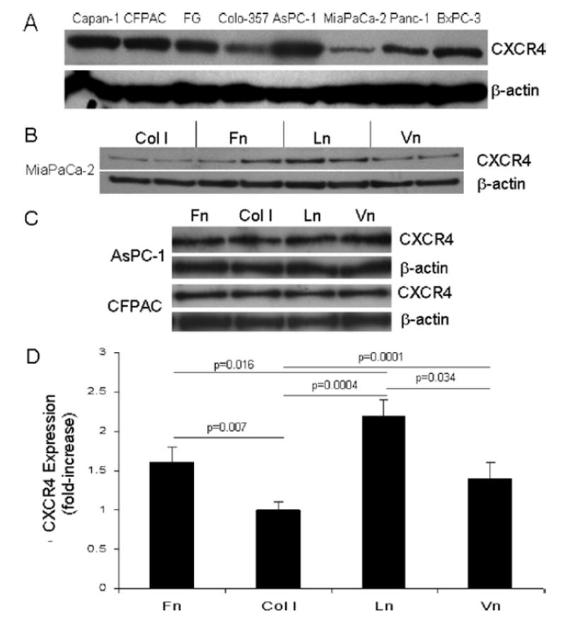
Background: We have shown recently that alpha(2)beta(1) integrin-mediated type I collagen adhesion promotes a more malignant phenotype in pancreatic cancer cell lines than other extracellular matrix (ECM) proteins. MiaPaCa-2 cells, by contrast, do not express collagen-binding integrins, but are metastatic in our orthotopic mouse model and migrate maximally on laminin-1 (Ln-1). It has also been shown that CXCR4 and IL-8 expression correlates directly with metastasis in pancreatic cancer in vivo. We therefore examined the potential of the ECM to regulate CXCR4 and IL-8 expression in pancreatic cancer cells.

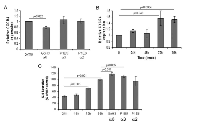
Methods: We cultured 8 pancreatic cancer cell lines on fibronectin (Fn), types I and IV collagen, Ln-1 and vitronectin (Vn), and examined cell lysates for CXCR4 by immunoblotting and media for IL-8 by ELISA. We also conducted cell migration assays with stromal-derived factor-1 (SDF-1) as the chemoattractant to examine integrin-binding specificity and CXCR4 function.

Results: All cell lines expressed CXCR4 protein. MiaPaCa-2 cell growth on Ln-1 increased significantly CXCR4 and IL-8 expression relative to other ECM proteins. Migration inhibition studies showed that both the alpha(6)beta(1) and alpha(3)beta(1) integrins mediate MiaPaCa-2 migration on Ln-1. Growth studies showed further that CXCR4 expression on Ln-1 was mediated by the alpha(6)beta(1) integrin whereas IL-8 expression was mediated by both the alpha(6)beta(1) and alpha(3)beta(1) integrins. The expression of functional CXCR4 was also shown in migration assays, where SDF-1 significantly increased pancreatic cancer cell chemotaxis on Ln-1.

Conclusions: These data indicate that integrin-mediated Ln-1 adhesion upregulates CXCR4 and IL-8 expression and may play a mechanistic role in pancreatic cancer metastases.


Figures

Fig 1
Fig 1
Pancreatic cancer cell lines express the CXCR4 chemokine receptor, and the ECM differentially regulates CXCR4 expression in MiaPaCa-2 pancreatic cancer cells. A, The indicated pancreatic cancer cell lines were grown on tissue culture plastic in DMEM supplemented with 10% FBS, lysates were prepared, and total protein determined as described in Materials and Methods. Ten micrograms of total protein were separated subsequently on 10% NuPage gels under reducing conditions and transferred to nitrocellulose using standard electrophoretic techniques. Membranes were exposed to polyclonal antisera (Clone G19; Santa Cruz Biotechnology) raised against CXCR4, followed by horseradish peroxidase-conjugated secondary antibody. The peroxidase was then detected using chemiluminescence according to manufacturer’s instructions (ECL kit; Amersham), and autoradiography. Membranes were also exposed to a monoclonal anti-β-actin antibody (Clone AC-15; Sigma), followed by horseradish peroxidase-conjugated secondary (Jackson ImmunoResearch Labs), and developed as described above. B, MiaPaCa-2 cells were cultured for 96 hours on Fn, type I collagen, Ln-1, or Vn, and total cell lysates were analyzed subsequently by immunoblotting for CXCR4 expression as described in Materials and Methods. β-actin immunoblotting results are also shown as a control for protein loading and for subsequent densitometry analyses. C, AsPC-1 and CFPAC cells were cultured for 96 hours on the indicated ECM proteins and analyzed by immunoblotting for CXCR4 expression as described. D, Immunoblotting results described in (B) above for CXCR4 expression in MiaPaCa-2 cells from 2 to 5 independent experiments conducted in triplicate for each substrate were analyzed by densitometry, and are expressed as fold-increase over type I collagen, which was arbitrarily assigned a value of 1. Significant differences in CXCR4 expression were observed between all substrates as indicated except in comparisons between Fn and Vn.
Fig 2
Fig 2
ECM regulates the expression of IL-8 in pancreatic cancer cell lines. MiaPaCa-2, AsPC-1, CFPAC, and BxPC-3 cells were cultured for 96 hours on Fn, type I collagen, Ln-1, or Vn, under serum-free conditions as described in Materials and Methods, and conditioned media were subsequently analyzed by ELISA for IL-8 expression. Results presented were normalized to total cellular protein, expressed as % Max for each cell line, and represent the mean ± SEM from 2 to 5 experiments with duplicate determinations. Statistically significant differences were noted in MiaPaCa-2 and AsPC-1 cells in comparisons between all substrates except Fn and Vn as indicated. Significant differences were also noted in CFPAC cells as indicated between Fn and both Ln and Vn, and between type I collagen and Ln. No significant differences were noted in BxPC-3 cells.
Fig 3
Fig 3
The α6β1 and α3β1 integrins mediate MiaPaCa-2 haptokinesis on Ln-1 substrates. Filters (8-μm pores) were coated with Ln-1 at 5 μg/ml and 5 × 10⁴ MiaPaCa-2 cells/well were examined for haptokinetic migration under serum-free conditions using the modified Boyden chamber in the presence or absence of 25 μg/ml anti-integrin function-blocking monoclonal antibodies as described. Data presented are the mean ± SEM from 3 experiments, with 6 replicates per treatment per experiment. Significant differences compared with untreated control wells are indicated.
Fig 4
Fig 4
The α6β1 integrin regulates CXCR4 and both the α6β1 and α3β1 integrins regulate IL-8 expression on Ln-1 substrates in MiaPaCa-2 cells. A, MiaPaCa-2 cells were cultured on Ln-1 substrates for 96 hours under serum-free conditions in the presence or absence of anti-integrin function blocking monoclonal antibodies directed against the α6, α3, and α2 subunits at 25 μg/ml as described in Materials and Methods. Lysates were then prepared, protein was determined, and 3 μg total protein per treatment group were examined for CXCR4 expression by immunoblotting and densitometry as described in Fig 1 and Materials and Methods. Densitometry data presented represent the mean ± SEM from two independent experiments conducted in duplicate for each treatment group. The antibody clones and the integrin subunits they are directed against are shown on the x-axis. Significant differences from the 96-hour untreated control cultures are indicated. B, MiaPaCa-2 cells were grown on Ln-1 substrates as described in Materials and Methods over a 96-hour time course, and harvested at the indicated time points. Cell lysates were prepared and analyzed by immunoblotting and subsequent densitometry for CXCR4 expression. Densitometry results represent the mean ± SEM from 2 independent experiments with duplicate wells per time point per experiment. Significant differences from the tissue culture plastic controls (T = 0) harvested at the time of initial cell seeding are indicated. C, Conditioned culture media from MiaPaCa-2 cells grown on Ln-1 over a 96-hour time course as described above were analyzed by ELISA for IL-8 expression. Results presented represent the mean ± SEM from 2 independent experiments conducted in duplicate. Statistically significant differences in comparisons to 24-hour IL-8 expression levels are indicated, as are significant differences in cells treated with monoclonal antibodies compared with 96-hour untreated control cultures.
Fig 5
Fig 5
SDF-1 increases pancreatic cancer cell migration on Ln-1 substrates in a dose-dependent manner. A, Chemotactic migration assays were conducted on Ln-1 substrates under serum-free conditions using modified Boyden chambers in the presence or absence of SDF-1 (100 ng/ml) as the chemoattractant as described in Materials and Methods. MiaPaCa-2, FG, BxPC-3, CFPAC, or AsPC-1 cells were added to the upper chambers at 5 × 10⁴/well and the entire apparatus was incubated at 37°C for 24 hours. Data presented are expressed as % Max, with BxPC-3 exhibiting maximal migration, and represent the mean ± SEM from 2 independent experiments with 4 replicates per treatment group. Black bars, with SDF-1; gray bars, without SDF-1. Significant differences are indicated. B, An SDF-1 dose-response curve was generated using chemotaxis assays as described above and in Materials and Methods with MiaPaCa-2 cells on Ln-1 substrates. The SDF-1 concentrations are indicated on the x-axis. Data presented are expressed as % Max, and represent the mean ± SEM from 2 independent experiments with 6 replicates per treatment group per experiment.

Similar articles

Cited by

References

    1. Bardeesy N, DePinho RA. Pancreatic cancer biology and genetics. Nat Rev Cancer. 2002;2:897–909. - PubMed
    1. Ruoslahti E. RGD and other recognition sequences for integrins. Annu Rev Cell Dev Biol. 1996;12:697–715. - PubMed
    1. Weinel RJ, Rosendahl A, Neumann K, Chaloupka B, Erb D, Rothmund M, et al. Expression and function of VLA-alpha 2, -alpha 3, -alpha 5 and -alpha 6-integrin receptors in pancreatic carcinoma. Int J Cancer. 1992;52:827–33. - PubMed
    1. Rosendahl A, Neumann K, Chaloupka B, Rothmund M, Weinel RJ. Expression and distribution of VLA receptors in the pancreas: an immunohistochemical study. Pancreas. 1993;8:711–8. - PubMed
    1. Shimoyama S, Gansauge F, Gansauge S, Oohara T, Beger HG. Altered expression of extracellular matrix molecules and their receptors in chronic pancreatitis and pancreatic adenocarcinoma in comparison with normal pancreas. Int J Pancreatol. 1995;18:227–34. - PubMed

Publication types

MeSH terms