. 2007 Jun 12;49(23):2292-300.
doi: 10.1016/j.jacc.2007.02.050. Epub 2007 May 25.

An elastic, biodegradable cardiac patch induces contractile smooth muscle and improves cardiac remodeling and function in subacute myocardial infarction

Affiliations

An elastic, biodegradable cardiac patch induces contractile smooth muscle and improves cardiac remodeling and function in subacute myocardial infarction

Kazuro L Fujimoto et al. J Am Coll Cardiol. .

Abstract

Objectives: Our objective in this study was to apply an elastic, biodegradable polyester urethane urea (PEUU) cardiac patch onto subacute infarcts and to examine the resulting cardiac ventricular remodeling and performance.

Background: Myocardial infarction induces loss of contractile mass and scar formation resulting in adverse left ventricular (LV) remodeling and subsequent severe dysfunction.

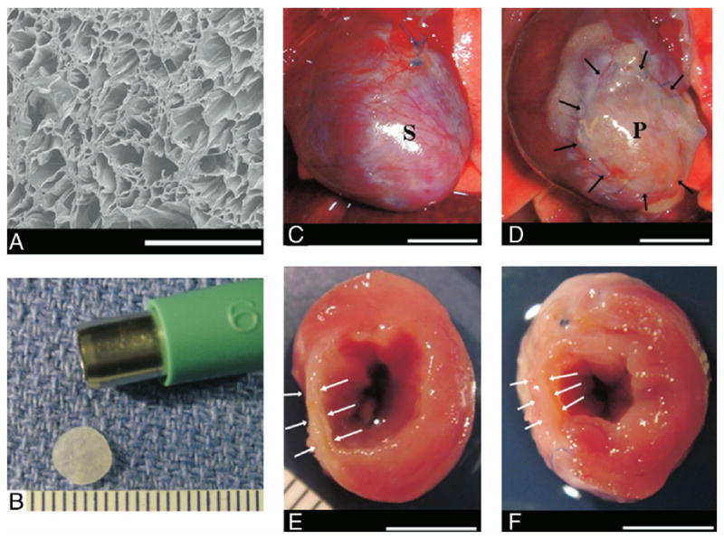
Methods: Lewis rats underwent proximal left coronary ligation. Two weeks after coronary ligation, a 6-mm diameter microporous PEUU patch was implanted directly on the infarcted LV wall surface (PEUU patch group, n = 14). Sham surgery was performed as an infarction control (n = 12). The LV contractile function, regional myocardial wall compliance, and tissue histology were assessed 8 weeks after patch implantation.

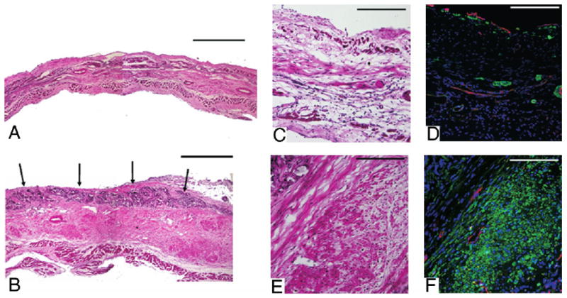
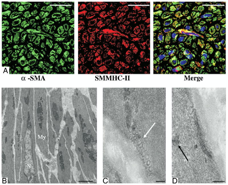
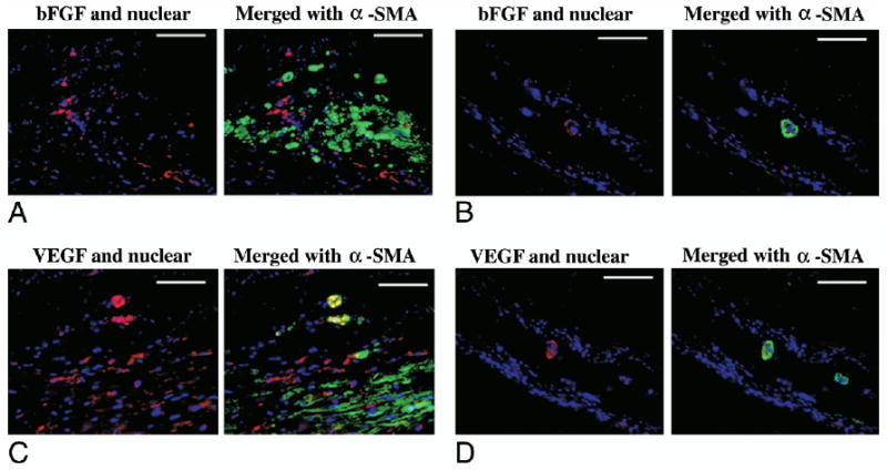
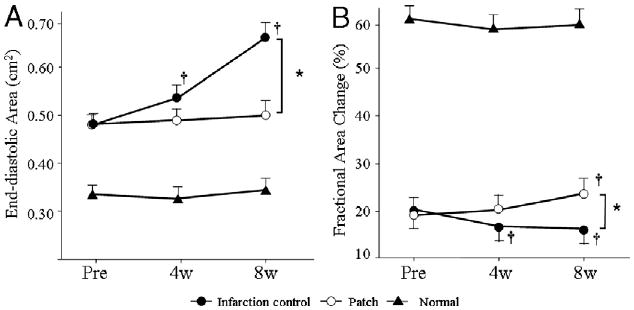
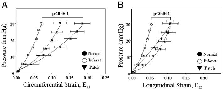
Results: The end-diastolic LV cavity area (EDA) did not change, and the fractional area change (FAC) increased in the PEUU patch group (p < 0.05 vs. week 0), while EDA increased and FAC decreased in the infarction control group (p < 0.05). The PEUU patch was largely resorbed 8 weeks after implantation and the LV wall was thicker than infarction control (p < 0.05 vs. control group). Abundant smooth muscle bundles with mature contractile phenotype were found in the infarcted myocardium of the PEUU group. The myocardial compliance of the PEUU group was distributed between normal myocardium and infarction control (p < 0.001).

Conclusions: Implantation of a novel biodegradable PEUU patch onto a subacute myocardial infarction promoted contractile phenotype smooth muscle tissue formation and improved cardiac remodeling and contractile function at the chronic stage. Our findings suggest a new therapeutic option against post-infarct cardiac failure.


Figures

Figure 1
Figure 1. Features of PEUU and Representative Images, at 8 Weeks After Implantation
Electron micrograph of the polyester urethane urea (PEUU) material (A). Polyester urethane urea patch in final format (6-mm diameter × 300 μm thick) (B). Representative images, 8 weeks after implantation, of the anterior view of infarction control (C) and PEUU patched (D) hearts. The cross-sectional view of both groups is shown in E and F, respectively. Black arrows point to the implanted PEUU patch, and white arrows indicate the infarcted anterior wall. Scale bar: 500 μm in A, 1 mm in B, 55 mm in C to F. P = the patch implanted area; S = the infarcted scar.
Figure 2
Figure 2. Representative Histological Sections
Representative histological sections of the infarction control (A) and PEUU patched (B) myocardial wall 8 weeks after implantation stained with hematoxylin and eosin. Black arrows indicate the top of the PEUU implanted area, which appears dark violet. Higher magnification of hematoxylin and eosin staining and immunohistochemical staining appear in C to F where C and D are infarcted control and E and F are PEUU patched. α-SMA staining appears green, CD31 staining appears red, and nuclear staining appears blue. Increased smooth muscle actin is apparent in the PEUU patched group. Scale bars in A and B are 500 μm. For C to F, scale bars are 200 μm.
Figure 3
Figure 3. Characterization of α-SMA–Positive Cells
Smooth muscle myosin heavy chain isotype II (SMMHC-II) (A) immunohistochemical staining colocalized with alpha-smooth muscle actin (α-SMA)–positive cells suggesting mature contractile ability. Electron micrographs (B to D) of the muscle-like bundles beneath the polyester urethane urea patch exhibited ultrastructural features typical of mature contractile phenotype smooth muscle cells. White arrow denotes caveole. Black arrow indicates dense bodies. Scale bars for A are 20 μm, for B are 2 μm, and for C and D are 100 nm. My = myofibril; N = nuclear.
Figure 4
Figure 4. bFGF and VEGF Immunohistochemical Staining
Immunohistochemical staining for basic fibroblast growth factor (bFGF) (A and B) and vascular endothelial growth factor (VEGF) (C and D) in the polyester urethane urea patch (A and C) and infarction control (B and D) groups. The left image of each set shows growth factor and nuclear staining, and the right image merges in alpha-smooth muscle actin (α-SMA) staining. Growth factor staining is red, α-SMA staining is green, and nuclear staining is blue. Scale bars are 100 μm.
Figure 5
Figure 5. Echocardiographic Assessment
Echocardiographic assessment of the polyester urethane urea patch, infarction control, and normal control groups during the study period. End-diastolic area is shown in (A), and % fractional area change in (B). End-diastolic area and % fractional area change in the normal control group were significantly different than those in the patch and infarction control groups (p < 0.05) at each time point and did not significantly vary during follow-up. *p < 0.05 between groups; †p < 0.05 versus just before implantation (pre) within group. 4w = 4 weeks after implantation; 8w = 8 weeks after implantation.
Figure 6
Figure 6. Pressure-Strain Relationship for Biomechanical Compliance
Pressure-strain relationship for biomechanical compliance of the left ventricular anterior wall. Circumferential strain (E11) is shown in (A) and longitudinal strain (E22) in (B). For both directions, the infarction control group exhibited the least compliance, while the polyester urethane urea patch group was significantly more compliant, being between infarcted and normal or equivalent to normal tissue.

Similar articles

Cited by

References

    1. Mann DL, Bristow MR. Mechanisms and models in heart failure: the biomechanical model and beyond. Circulation. 2005;111:2837–49. - PubMed
    1. Holmes JW, Borg TK, Covell JW. Structure and mechanics of healing myocardial infarcts. Annu Rev Biomed Eng. 2005;7:223–53. - PubMed
    1. White HD, Norris RM, Brown MA, Brandt PW, Whitlock PM, Wild CJ. Left ventricular end-systolic volume as the major predictor of survival after recovery from myocardial infarction. Circulation. 1987;76:44–51. - PubMed
    1. Sabbah HN, Shimoyama H, Kono T, et al. Effects of long-term monotherapy with enalapril, metoprolol, and digoxin on the progression of left ventricular dysfunction and dilation in dogs with reduced ejection fraction. Circulation. 1994;89:2852–9. - PubMed
    1. Dor V, Sabatier M, DiDonato M, Montiglio F, Toso A, Maioli M. Efficacy of endoventricular patch plasty in large postinfarction akinetic scar and severe left ventricular dysfunction: comparison with a series of large dyskinetic scars. J Thorac Cardiovasc Surg. 1998;116:50–9. - PubMed

Publication types

MeSH terms