. 2007 Aug 1;74(3):425-37.
doi: 10.1016/j.bcp.2007.04.024. Epub 2007 May 3.

Inhibition of the HERG potassium channel by the tricyclic antidepressant doxepin

Affiliations

Inhibition of the HERG potassium channel by the tricyclic antidepressant doxepin

R S Duncan et al. Biochem Pharmacol. .

Abstract

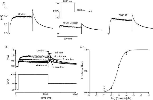
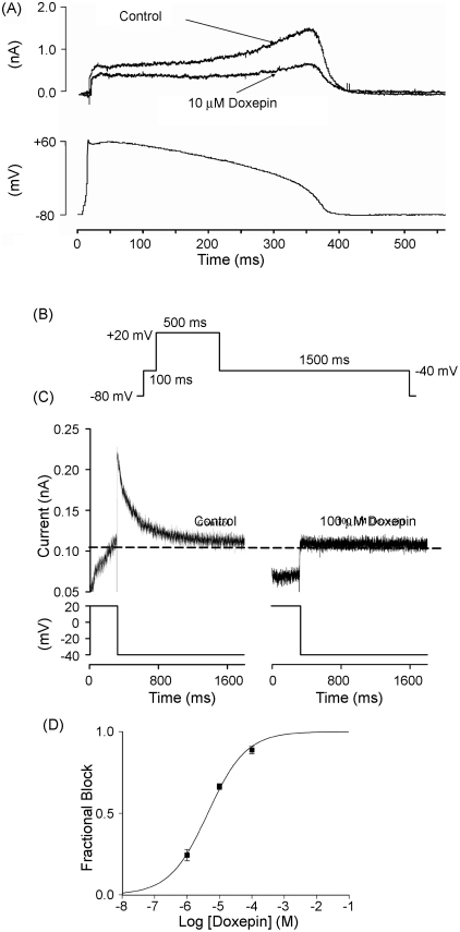
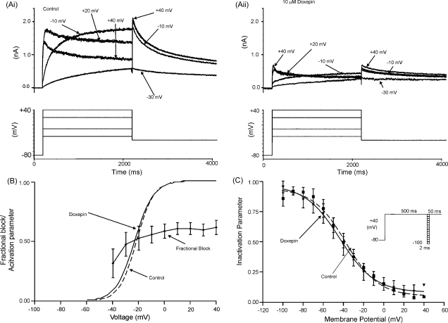
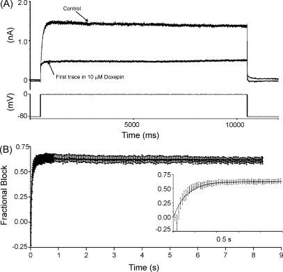
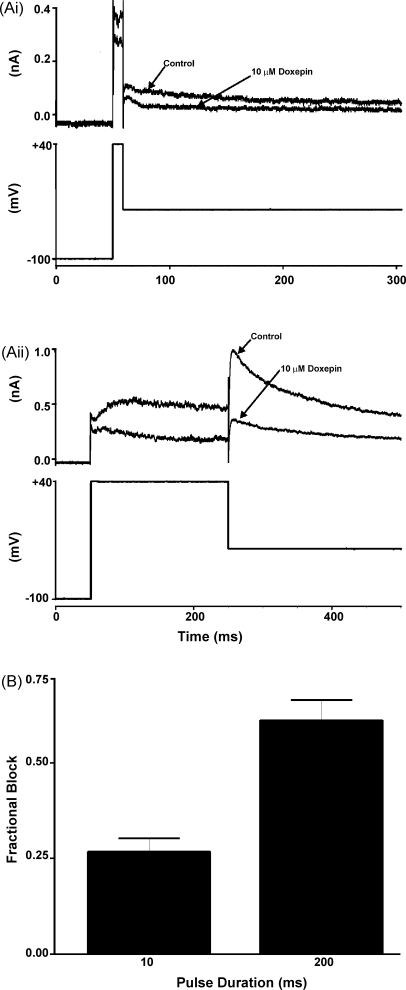
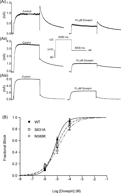
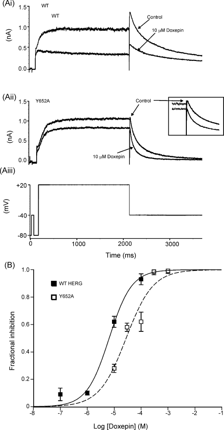
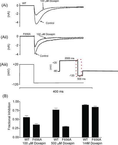
HERG (human ether-à-go-go-related gene) encodes channels responsible for the cardiac rapid delayed rectifier potassium current, I(Kr). This study investigated the effects on HERG channels of doxepin, a tricyclic antidepressant linked to QT interval prolongation and cardiac arrhythmia. Whole-cell patch-clamp recordings were made at 37 degrees C of recombinant HERG channel current (I(HERG)), and of native I(Kr) 'tails' from rabbit ventricular myocytes. Doxepin inhibited I(HERG) with an IC(50) value of 6.5+/-1.4 microM and native I(Kr) with an IC(50) of 4.4+/-0.6 microM. The inhibitory effect on I(HERG) developed rapidly upon membrane depolarization, but with no significant dependence on voltage and with little alteration to the voltage-dependent kinetics of I(HERG). Neither the S631A nor N588K inactivation-attenuating mutations (of residues located in the channel pore and external S5-Pore linker, respectively) significantly reduced the potency of inhibition. The S6 point mutation Y652A increased the IC(50) for I(HERG) blockade by approximately 4.2-fold; the F656A mutant also attenuated doxepin's action at some concentrations. HERG channel blockade is likely to underpin reported cases of QT interval prolongation with doxepin. Notably, this study also establishes doxepin as an effective inhibitor of mutant (N588K) HERG channels responsible for variant 1 of the short QT syndrome.


Figures

Fig. 1
Fig. 1
Concentration dependent inhibition of IHERG by doxepin. (A) Representative currents in the absence, presence (5 min) and following wash off (5 min) of 10 μM doxepin, elicited by the voltage protocol shown in the inset. (B) Representative currents (upper traces; lower trace shows voltage protocol), from the same cell as in A, in the absence and during doxepin exposure. ‘Control’ refers to current prior to doxepin exposure, whilst numbered time-points indicate currents sampled at 1–5 min of doxepin application. (C) Mean fractional block produced by four different concentrations of doxepin fitted with Eq. (2), which yielded an IC50 value of 6.5 μM (± 1.4 μM) with a Hill coefficient of 1.0 (± 0.2). Each drug concentration was applied to a minimum of five cells.
Fig. 2
Fig. 2
Doxepin inhibition of IHERG during action potential (AP) voltage clamp and of native IKr tails. (A) The upper traces show (leak corrected) representative currents of heterologously expressed IHERG in the absence and presence of 10 μM doxepin elicited by an AP clamp protocol (lower trace; voltage command applied at 4 s intervals). (B) Schematic representation of protocol used to assess blockade of native IKr tails by doxepin. (C) Representative tail currents from a rabbit ventricular myocyte upon repolarization from +20 to −40 mV in the absence (left panel) and in the presence of 100 μM doxepin (right panel). Upper traces show current records and lower traces show corresponding portion of the voltage protocol. The horizontal dashed line is drawn at the level of the current at −40 mV at the end of the initial (100 ms) step to −40 mV, against which peak IKr amplitude on repolarization from +20 to +40 mV was measured. (D) Mean fractional tail current block produced by three different concentrations of doxepin fitted with Eq. (2), which yielded an IC50 value of 4.4 (± 0.6 μM) with a Hill coefficient of 0.7 (± 0.1). Each drug concentration was applied to a minimum of twelve cells.
Fig. 3
Fig. 3
Voltage dependence of IHERG inhibition by doxepin. (A) Representative currents (upper traces) at selected voltages in: (Ai) the absence and (Aii) presence of 10 μM doxepin, elicited by the voltage-protocol shown in the lower traces (only some steps shown, for purposes of clarity of display). (B) Mean fractional block (n = 5) of peak IHERG tails produced by 10 μM doxepin, between test potentials of −40 and +40 mV. Superimposed are the continuous plots of activation curves for IHERG in the absence (dashed line) and presence (continuous line) of doxepin, calculated as described in Section 2. The mean activation V0.5 and k values obtained from IHERG tail-voltage relations using Eq. (3) are given in Section 3. (C) Voltage-dependence of IHERG availability/inactivation (protocol shown in inset). The mean (n = 5) normalised data (I/Imax) were plotted against the test potential and fitted with Eq. (4). The derived half-maximal inactivation values were: control V0.5 = −37.9 ± 2.4 mV and doxepin V0.5 = − 43.5 ± 4.1 mV in the absence (filled squares, dashed line) and presence (filled triangles, continuous line) of 10 μM doxepin, respectively (ns paired t-test p = 0.79), with corresponding k values of −16.2 ± 2.4 and −17.3 ± 8.8 mV (ns paired t-test p = 0.59).
Fig. 4
Fig. 4
Time-dependence of development of IHERG inhibition during a sustained depolarization. (A) Lower trace shows voltage-protocol (10 s depolarizing step from −80 to 0 mV) used to elicit IHERG in the absence of doxepin and following a 7-min exposure to 10 μM doxepin in the absence of pulsing. (B) Mean fractional block of IHERG (n = 8) produced by 10 μM doxepin during the 10-s depolarizing step to 0 mV, following 7-min of drug application in the absence of pulsing. Inset shows, on an expanded time-scale, the development of blockade over the first 500 ms of the protocol. Monoexponential fitting of the fractional block time relationship yielded a rate constant (K) for the fit of 16.33 ± 0.73 s−1 (equivalent to a time-constant (1/K) of 61 ms).
Fig. 5
Fig. 5
IHERG inhibition by doxepin with brief-duration voltage-commands. (A) For each of (Ai) and (Aii), the upper traces show IHERG elicited by the voltage protocol shown in the lower trace: (Ai) shows representative traces of currents elicited by a 10-ms duration voltage command from −100 to +40 mV in the absence and presence of 10 μM doxepin; (Aii) shows data from the same cell with a 200 ms duration command. (B) Plot of mean (±S.E.M.) fractional block of IHERG tails following the 2 different duration commands (n = 5; p < 0.02).
Fig. 6
Fig. 6
(A) Effect of inactivation mutants on IHERG inhibition by doxepin. (Ai) Representative WT-HERG. (Aii) S631A-HERG and (Aiii) N588K-HERG currents in the absence (control, left hand traces) and presence of 10 μM doxepin (right hand traces). The voltage protocol is shown as an inset. (B). Mean fraction block of IHERG by 1, 10 and 100 μM doxepin (each drug concentration was applied to at least five cells; data for WT IHERG replotted from Fig. 1) fitted with Eq. (2). The derived IC50 values for WT IHERG, S631A–HERG and N588K–HERG were, respectively, 6.6 ± 0.6, 8.6 ± 0.0 and 12.6 ± 2.7 μM (p > 0.05; Anova); Hill coefficients for the fits to WT, S631A and N588K data were, respectively, 1.1 ± 0.1, 0.8 ± 0.0, and 0.7 ± 0.l (p < 0.05 between WT and N588K).
Fig. 7
Fig. 7
(A) Effect of the Y652A mutation on IHERG inhibition by doxepin. (Ai) Representative WT-HERG and (Aii) Y652A-HERG currents in the absence and (Aiii) the presence of 10 μM doxepin, elicited by voltage protocol. Inset in (Aii) shows expanded tail-currents to highlight attenuation of blockade for the Y652A mutant.(B) Concentration-response relations for inhibition of IHERG by doxepin for WT–HERG and Y652A–HERG, fitted with Eq. (2). Data for WT IHERG are replotted from Fig. 1. For Y652A–HERG the following concentrations were tested: 10 μM (n = 4), 30 μM (n = 4); 100 μM (n = 12); 300 μM (n = 4) and 1 mM (n = 4). The derived IC50 values for WT IHERG and Y652A IHERG were, respectively, 6.5 ± 1.4 and 27.8 ± 8.8 μM (p < 0.05); Hill coefficients for the fits to WT IHERG and Y652A IHERG of 1.0 ± 0.2 and 0.9 ± 0.3 (p > 0.05).
Fig. 8
Fig. 8
(A) Effect of the F656A mutation on IHERG inhibition by doxepin. (Ai) Representative WT–HERG and (Aii) F656A-HERG currents in the absence and presence of 100 μM doxepin, elicited by voltage protocol (Aiii) on an expanded time-scale (full protocol shown as an inset). (B) Bar chart showing the mean fractional block levels produced for WT–HERG and F656A–HERG tails measured at −120 mV after addition of 100, 500 μM or 1 mM doxepin (n = 5 for each). The F656A mutation significantly attenuated the level of blockade produced by both 100 μM doxepin (p < 0.01) and 500 μM doxepin (p < < 0.001), but not that by 1 mM doxepin (p > 0.05).

Similar articles

Cited by

References

    1. Viskin S. Long QT syndromes and torsade de pointes. Lancet. 1999;354:1625–1633. - PubMed
    1. Haverkamp W., Breithardt G., Camm A.J., Janse M.J., Rosen M.R., Antzelevitch C. The potential for QT prolongation and pro-arrhythmia by non-anti-arrhythmic drugs: clinical and regulatory implications report on a policy conference of the European Society of Cardiology. Cardiovasc Res. 2000;47:219–233. - PubMed
    1. Shah R.R. Drugs QT interval prolongation and ICH E14—the need to get it right. Drug Safety. 2005;28:115–125. - PubMed
    1. Witchel H.J., Hancox J.C. Familial and acquired long QT syndrome and the cardiac rapid delayed rectifier potassium current. Clin Exp Pharmacol Physiol. 2000;27:753–766. - PubMed
    1. Vandenberg J.I., Walker B.D., Campbell T.J. HERG K+ channels: friend and foe. Trends Pharmacol Sci. 2001;22:240–246. - PubMed

Publication types

MeSH terms