. 2007 Aug 3;371(1):210-21.
doi: 10.1016/j.jmb.2007.05.038. Epub 2007 May 18.

A novel loop domain in superantigens extends their T cell receptor recognition site

Affiliations

A novel loop domain in superantigens extends their T cell receptor recognition site

Sebastian Günther et al. J Mol Biol. .

Abstract

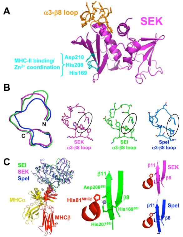
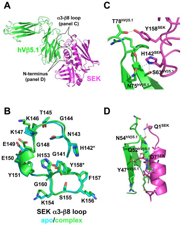
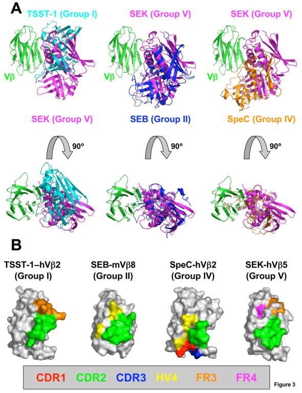
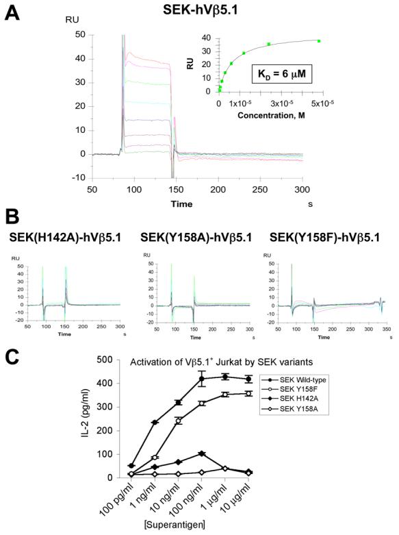
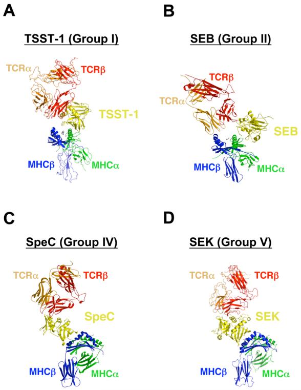
Superantigens (SAGs) interact with host immune receptors to induce a massive release of inflammatory cytokines that can lead to toxic shock syndrome and death. Bacterial SAGs can be classified into five distinct evolutionary groups. Group V SAGs are characterized by the alpha3-beta8 loop, a unique approximately 15 amino acid residue extension that is required for optimal T cell activation. Here, we report the X-ray crystal structures of the group V SAG staphylococcal enterotoxin K (SEK) alone and in complex with the TCR hVbeta5.1 domain. SEK adopts a unique TCR binding orientation relative to other SAG-TCR complexes, which results in the alpha3-beta8 loop contacting the apical loop of framework region 4, thereby extending the known TCR recognition site of SAGs. These interactions are absolutely required for TCR binding and T cell activation by SEK, and dictate the TCR Vbeta domain specificity of SEK and other group V SAGs.


Figures

Figure 1
Figure 1
Structural similarity of SEK and other Group V bacterial superantigens. (A) Structure of SEK in the unbound state. The α3-β8 loop is in orange and the residues responsible for Zn2+ coordination and MHC class II binding are in cyan. (B) Structural comparison of the α3-β8 loop domains of SEK, SEI and SpeI. Superposition of the main chains of the α3-β8 loops (left panel). Molecular detail of side chain positions in the α3-β8 loops (right panels). SEK, SEI and SpeI are in magenta, green and blue, respectively. The residues in SEK that interact with TCR, His142 and Tyr158, as well as corresponding residues in SEI and SpeI are encircled. (C) Structural comparison of the MHC binding site of SEI and the putative MHC binding sites of SEK and SpeI. Superposition of SEK and SpeI with SEI from the SEI-MHC class II crystal structure (left panel). Close-up view of the Zn2+ coordination between SEI residues His169, His207 and Asp209 and the MHC β subunit residue His81 (middle panel). Close-up views of the putative SAG-MHC interface formed by the superposed SEK and SpeI structures (right panels). The MHC α subunit is in yellow, MHC β subunit is in red, zinc ion in gray, and the SAG colors are as in panel B.
Figure 2
Figure 2
Structure of the SEK-hVβ5.1 complex. (A) Cartoon representation of the interaction between SEK and hVβ5.1. SEK is in magenta, hVβ5.1 is in green. The two regions of SEK, the α3-β8 loop and the N-terminus, that contact the TCR are encircled. (B) Comparison of SEK α3-β8 loop structures in the unbound (cyan) and hVβ5.1-bound (green) crystal structures. The residues that make intermolecular contacts with hVβ5.1, His142 and Tyr158, are demarcated by asterisks. (C) Close-up view of the interface formed by the α3-β8 loop of SEK and the FR3 and FR4 loops of hVβ5.1. (D) Close-up view of the interface formed by the N-terminus of SEK and the CDR2 loop of hVβ5.1. In both panels C and D, the side chains of only those residues that make intermolecular contacts are shown and side chain to side chain hydrogen bonds are shown as dashed lines.
Figure 3
Figure 3
Diverse TCR engagement by bacterial SAGs. (A) Superposition of the SEK-hVβ5.1 crystal structure with the TSST-1–hVβ2.1 (left panels), SEB-mVβ8.2 (middle panels) and SpeC-hVβ2.1 (right panels) complexes. The hVβ2.1 and mVβ8.2 molecules have been removed for clarity. SEK is in magenta, TSST-1 is in cyan, SEB is in blue, SpeC is in orange and Vβ is in green. (B) TCR Vβ domain molecular surface buried by various SAGs. Hypervariable and framework region surface residues buried in the interface formed by TSST-1, SEB, SpeC and SEK are color-coded as follows: CDR1 (red); CDR2 (green); CDR3 (blue); HV4 (yellow); FR3 (orange); and FR4 (magenta). The total buried surface areas for each SAG-TCR complex are: 1917 Å2 (TSST-1–hVβ2.1); 1268 Å2 (SEB-mVβ8.2); 1818 Å2 (SpeC-hVβ2.1); and 1572 Å2 (SEK-hVβ5.1).
Figure 4
Figure 4
Analysis of SEK-hVβ5.1 binding and SEK-mediated T cell activation. (A) SPR analysis of the wild type SEK-hVβ5.1 interaction. Non-linear regression analysis of maximal responses versus concentration is shown in the inset plot. (B) SPR analyses of hVβ5.1 interactions with the SEK(H142A), SEK(Y158A) and SEK(Y158F) mutants. (C) IL-2 secretion by eJRT3-5.1 cells incubated with various concentrations of wild type SEK and the SEK(H142A), SEK(Y158A) and SEK(Y158F) mutants presented by LG-2 cells. Data shown are the average ± SEM.
Figure 5
Figure 5
MHC-SAG-TCR ternary signaling complexes mediated by (A) TSST-1, (B) SEB, (C) SpeC, and (D) SEK. Colors are as follows: MHC α subunit, green; MHC β subunit, blue; antigenic peptide, gray; TCR α chain, orange; TCR β chain, red; SAGs, yellow. For clarity, the MHC-SAG-TCR complexes mediated by SpeC (panel C) and SEK (panel D) are rotated approximately 90 degrees clockwise about the vertical axis of the page relative to those mediated by TSST-1 (panel A) and SEB (panel B).

Similar articles

Cited by

References

    1. McCormick JK, Yarwood JM, Schlievert PM. Toxic shock syndrome and bacterial superantigens: an update. Annu Rev Microbiol. 2001;55:77–104. - PubMed
    1. Sundberg EJ, Li Y, Mariuzza RA. So many ways of getting in the way: diversity in the molecular architecture of superantigen-dependent T-cell signaling complexes. Curr Opin Immunol. 2002;14:36–44. - PubMed
    1. Kaul R, McGeer A, Norrby-Teglund A, Kotb M, Schwartz B, O'Rourke K, Talbot J, Low DE. Intravenous immunoglobulin therapy for streptococcal toxic shock syndrome--a comparative observational study. The Canadian Streptococcal Study Group. Clin Infect Dis. 1999;28:800–7. - PubMed
    1. LeClaire RD, Bavari S. Human antibodies to bacterial superantigens and their ability to inhibit T-cell activation and lethality. Antimicrob Agents Chemother. 2001;45:460–3. - PMC - PubMed
    1. Li Y, Li H, Dimasi N, McCormick JK, Martin R, Schuck P, Schlievert PM, Mariuzza RA. Crystal structure of a superantigen bound to the high-affinity, zinc-dependent site on MHC class II. Immunity. 2001;14:93–104. - PubMed

Publication types

MeSH terms

Associated data

LinkOut - more resources