Randomized Controlled Trial
. 2007 Oct;17(10):672-86.
doi: 10.1016/j.euroneuro.2007.04.002. Epub 2007 Jun 8.

The effect of doxapram on brain imaging in patients with panic disorder

Affiliations
Randomized Controlled Trial

The effect of doxapram on brain imaging in patients with panic disorder

Amir Garakani et al. Eur Neuropsychopharmacol. 2007 Oct.

Abstract

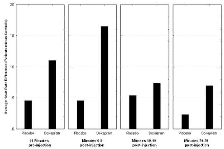
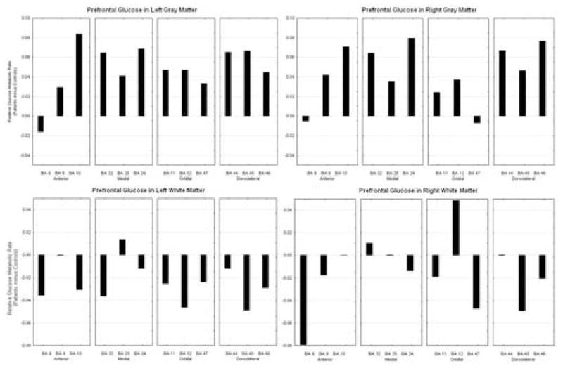
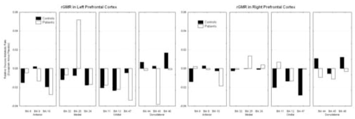
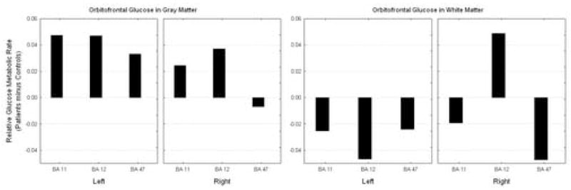
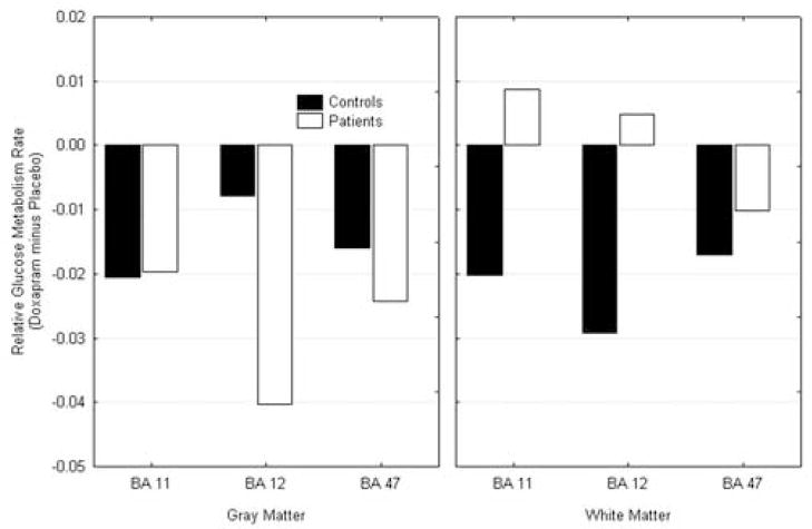
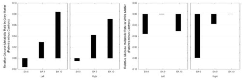
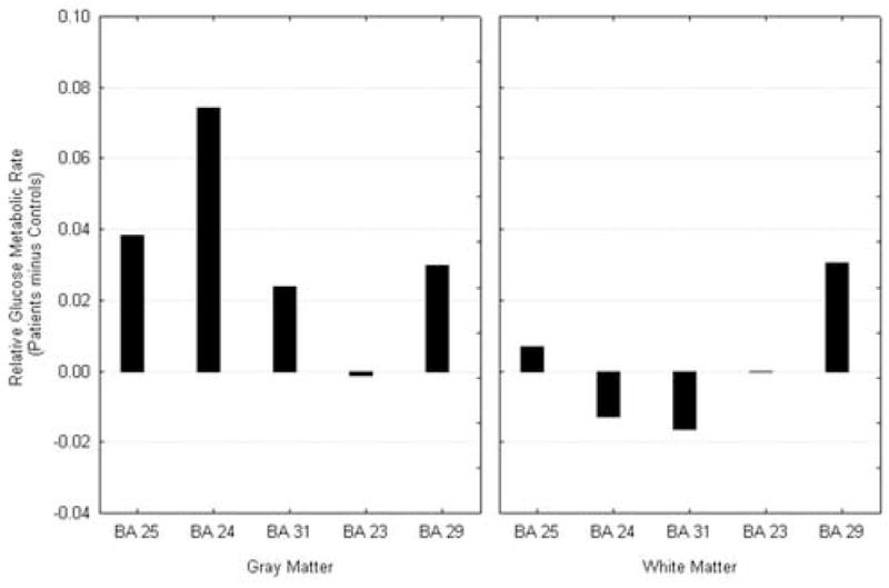
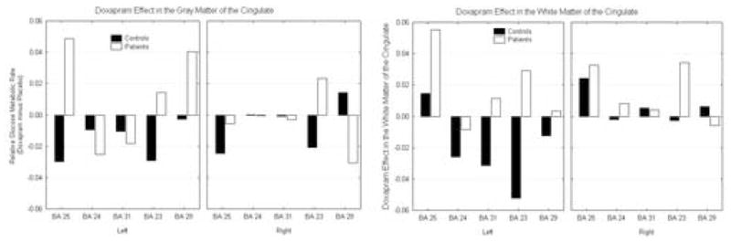
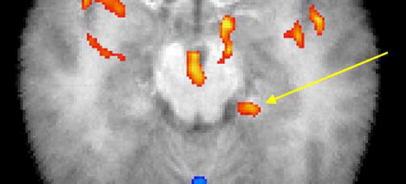
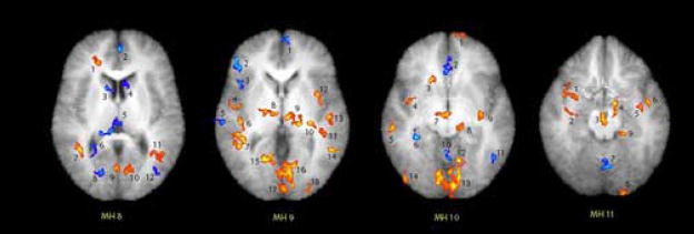
Administration of doxapram hydrochloride, a respiratory stimulant, is experienced by panic disorder patients to be similar to panic attacks but has reduced emotional effect in normal volunteers, thus providing a laboratory model of panic for functional imaging. Six panic patients and seven normal control subjects underwent positron emission tomography with (18)F-deoxyglucose imaging after a single-blinded administration of either doxapram or a placebo saline solution. Saline and doxapram were administered on separate days in counterbalanced order. Patients showed a greater heart rate increase on doxapram relative to saline than controls, indicating differential response. On the saline placebo day, patients had greater prefrontal relative activity than controls. In response to doxapram, patients tended to decrease prefrontal activity more than controls, and increased cingulate gyrus and amygdala activity more than controls. This suggests that panic disorder patients activate frontal inhibitory centers less than controls, a tendency that may lower the threshold for panic.


Figures

Figure 1
Figure 1
Effect of Doxapram on Heart Rate. Patient increase in heart rate relative to normal volunteer change (patient’s heart rate minus normal volunteer heart rate. Note patient heart rate is faster than controls but the increment in heart rate with doxapram is significantly larger. Post-hoc t-tests show no significant difference in 10-minutes pre-injection but significant in minutes 0–9 (normal volunteers 70.9, sd = 9.6, patients 87.3, sd = 5.65, t = 3.68, p = 0.0036).
Figure 2
Figure 2
On saline, patients generally show greater activity in the gray matter throughout the PFC, but decreased activity in the white matter.
Figure 3
Figure 3
Effect of doxapram on prefrontal cortex. Upon receiving doxapram, normals activate dorsolateral areas 44, 45, and 46, while patients show decreases in this area. In orbital and anterior cortex, both groups show decreases. In the medial frontal cortex, patients show a large inferior cingulate area 25 increase while both normals and patients show decreases in other areas.
Figure 4
Figure 4
Saline condition activity in the orbitofrontal cortex. Patients show increased activity in the orbitofrontal cortex gray matter and decreased activity in the white matter.
Figure 5
Figure 5
Doxapram effect in the orbitofrontal cortex. Both patients and controls showed deactivation in the gray matter of the orbitofrontal cortex with doxapram. The patients increased in activity in the white matter.
Figure 6
Figure 6
Saline condition activity in the anterior prefrontal cortex. In the anterior prefrontal cortex, the Panic Disorder patients show increased activity in the gray matter but decreased white matter activity.
Figure 7
Figure 7
Cingulate Activity at Saline condition. Cingulate gyrus arch shows patients with relatively higher activity than normals in the anterior portion and lower in the midsection; white matter shows decreases throughout.
Figure 8
Figure 8
Doxapram effect on the cingulate. Patients show increases and controls decreases in anterior cingulate gyrus gray matter and larger increases than controls in anterior cingulate white matter.
Figure 9
Figure 9
Statistical mapping for test of greater amygdala activation in patients with panic disorder than in normal controls. Confirmatory mapping of amygdala hypothesis: red indicates areas with greater doxapram-induced activation in panic disorder patients than in normal controls; t-test on doxapram minus placebo relative metabolic rate, p < 0.05, 1-tailed. Arrow marks 40 pixel patch in amygdala/hippocampus centered at −22, −13, −16; anterior edge at −22, −10, −16, which is on the edge of the gray matter of the parahippocampal gyrus 41%, amygdala 32%, 7×7×7 search box on Talairach daemon, using excel2td. http://ric.uthscsa.edu/projects/tdc/suggestedUses.html. The patch has a mean t of 2.36 and the highest t is 3.17, df = 11, p < 0.005.
Figure 10
Figure 10
SPM Maps of MH levels 8–11. Confirmatory mapping of amygdala hypothesis: red indicates areas with greater doxapram-induced activation in panic disorder patients than in normal controls; t-test on doxapram minus placebo relative metabolic rate. Each numbered patch refers to the table above. The threshold is p<0.05, 1-tailed (dark red margin of each spot), but the reader can choose the preferred p value from the table. Thus the center of the amygdala (see table) is 3.17, p = 0.005.

Similar articles

Cited by

References

    1. Abelson JL, Nesse RM, Weg JG, Curtis GC. Respiratory psychophysiology and anxiety: cognitive intervention in the doxapram model of panic. Psychosom Med. 1996a;58:302–313. - PubMed
    1. Abelson JL, Weg JG, Nesse RM, Curtis GC. Neuroendocrine responses to laboratory panic: cognitive intervention in the doxapram model. Psychoneuroendocrinology. 1996b;21:375–90. - PubMed
    1. Abelson JL, Liberzon I, Young EA, Khan S. Cognitive Modulation of the Endocrine Stress Response to a Pharmacological Challenge in Normal and Panic Disorder Subjects. Arch Gen Psychiatry. 2005;62:668–675. - PubMed
    1. Abelson JL, Khan S, Liberzon I, Young EA. HPA axis activity in patients with panic disorder: review and synthesis of four studies. Depress Anxiety. 2007;24:66–76. - PubMed
    1. Anand A, Shekhar A. Brain imaging studies in mood and anxiety disorders: special emphasis on the amygdala. Ann NY Acad Sci. 2003;985:370–388. - PubMed

Publication types