

Assessment of the canine model of rotator cuff injury and repair
- PMID: 17560802
- PMCID: PMC2204080
- DOI: 10.1016/j.jse.2007.04.002
Assessment of the canine model of rotator cuff injury and repair
Abstract
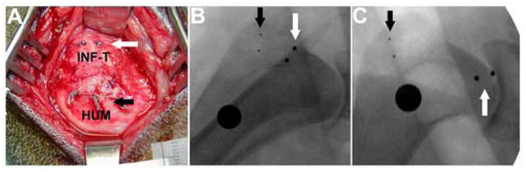
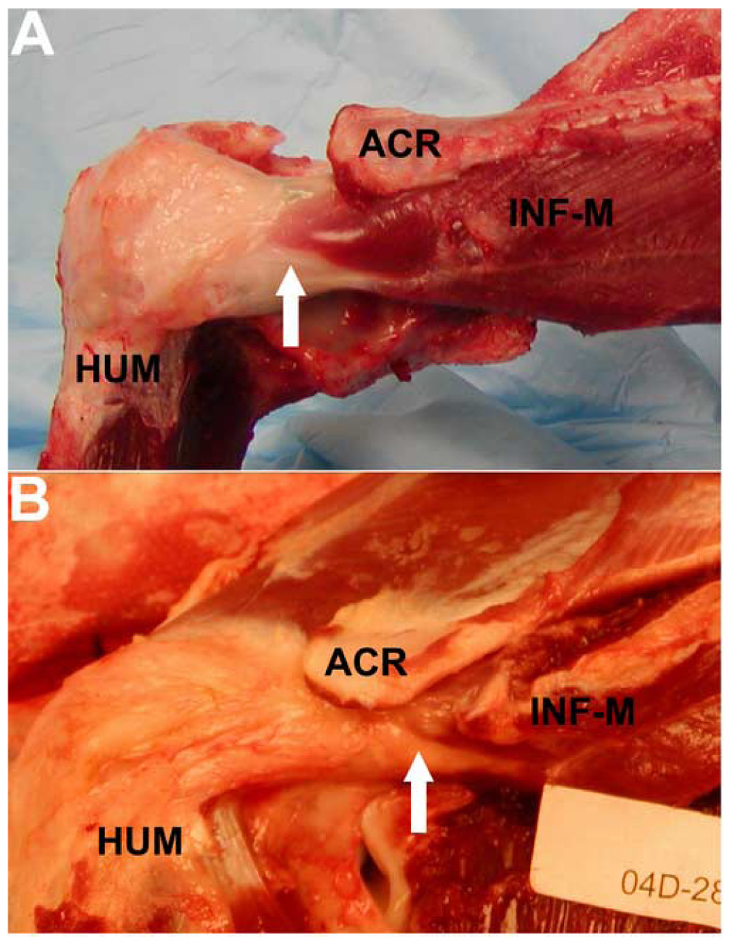
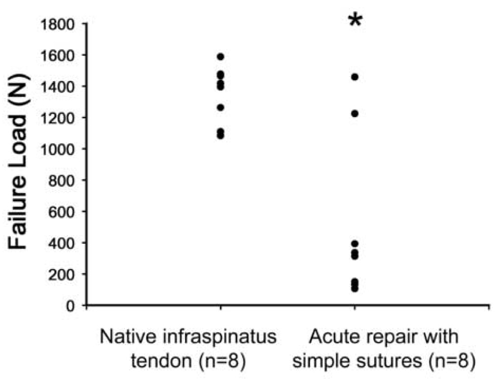
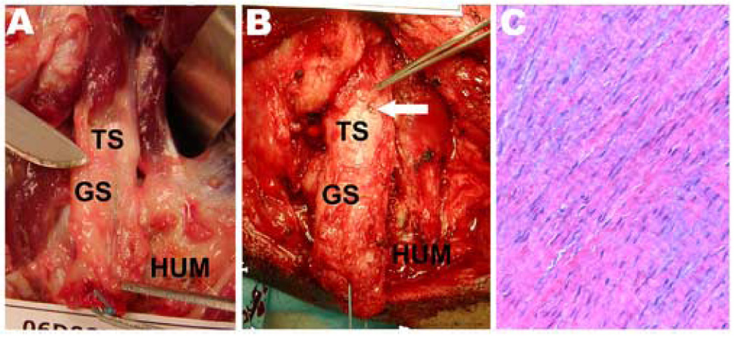
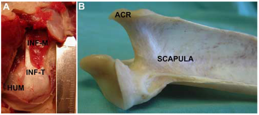
Animal shoulder models are used to systematically investigate the factors influencing rotator cuff injury and repair. Each model has advantages and disadvantages that must be considered in the context of the specific research questions being asked. This study evaluated the utility of the canine model for studies of acute, full-thickness rotator cuff tendon injury and repair. We found that time-zero failure load is dependent on the suture type and configuration used for repair. Acute, full-width tendon repairs fail anatomically within the first days after surgery in the canine model, regardless of suture type, suture configuration, or postoperative protocol. Robust scar tissue forms in the gap between the failed tendon end and the humerus, which can be visually, mechanically, and histologically misconstrued as tendon if an objective test of repair connectivity is not performed. We conclude that a full-width injury and repair model in the canine will provide a rigorous test of whether a new repair strategy or postoperative protocol, such as casting or temporary muscle paralysis, can maintain repair integrity in a high-load environment. Alternatively, a partial-width tendon injury model allows loads to be shared between the tendon repair and the remaining intact portion of the infraspinatus tendon and prohibits complete tendon retraction. Thus a partial-width injury in the canine may model the mechanical environment of many single tendon tears in the human injury condition and warrants further investigation.
Figures
References
-
- Adams JE, Zobitz ME, Reach JS, Jr, An KN, Steinmann SP. Rotator cuff repair using an acellular dermal matrix graft: an in vivo study in a canine model. Arthroscopy. 2006 Jul;22(7):700–709. - PubMed
-
- Anderst WJ, Les C, Tashman S. In vivo serial joint space measurements during dynamic loading in a canine model of osteoarthritis. Osteoarthritis Cartilage. 2005 Sep;13(9):808–816. - PubMed
-
- Aoki M, Miyamoto S, Okamura K, Yamashita T, Ikada Y, Matsuda S. Tensile properties and biological response of poly (L-lactic acid) felt graft: an experimental trial for rotator-cuff reconstruction. J Biomed Mater Res B Appl Biomater. 2004 Nov 15;71(2):252–259. - PubMed
-
- Aoki M, Oguma H, Fukushima S, Ishii S, Ohtani S, Murakami G. Fibrous connection to bone after immediate repair of the canine infraspinatus: the most effective bony surface for tendon attachment. J Shoulder Elbow Surg. 2001 Mar;10(2):123–128. - PubMed
-
- Bardet JF. Shoulder diseases in dogs. Vet Med. 2002;10:909–918.
Publication types
MeSH terms
Substances
Grants and funding
LinkOut - more resources
Full Text Sources
Other Literature Sources
Medical
Miscellaneous
